Liu Jin, Yin Ping, Wang Sicong, Liu Tao, Sun Chao, Hong Nan
Department of Radiology, Peking University People's Hospital, Beijing, China.
Pharmaceutical Diagnostic Team, GE Healthcare, Shanghai, China.
Front Oncol. 2021 Feb 26;11:628534. doi: 10.3389/fonc.2021.628534. eCollection 2021.
This study aims to assess the performance of radiomics approaches based on 3D computed tomography (CT), clinical and semantic features in predicting the pathological classification of thymic epithelial tumors (TETs).
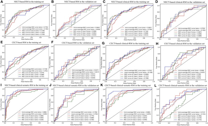
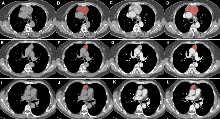
A total of 190 patients who underwent surgical resection and had pathologically confirmed TETs were enrolled in this retrospective study. All patients underwent non-contrast-enhanced CT (NECT) scans and contrast-enhanced CT (CECT) scans before treatment. A total of 396 hand-crafted radiomics features of each patient were extracted from the volume of interest in NECT and CECT images. We compared three clinical features and six semantic features (observed radiological traits) between patients with TETs. Two triple-classification radiomics models (RMs), two corresponding clinical RMs, and two corresponding clinical-semantic RMs were built to identify the types of the TETs. The area under the receiver operating characteristic curve (AUC) and accuracy (ACC) were useful to evaluate the different models.
Of the 190 patients, 83 had low-risk thymoma, 58 had high-risk thymoma, and 49 had thymic carcinoma. Clinical features (Age) and semantic features (mediastinal fat infiltration, mediastinal lymph node enlargement, and pleural effusion) were significantly different among the groups( < 0.001). In the validation set, the NECT-based clinical RM (AUC = 0.770 for low-risk thymoma, 0.689 for high-risk thymoma, and 0.783 for thymic carcinoma; ACC = 0.569) performed better than the CECT-based clinical-semantic RM (AUC = 0.785 for low-risk thymoma, 0.576 for high-risk thymoma, and 0.774 for thymic carcinoma; ACC = 0.483).
NECT-based and CECT-based RMs may provide a non-invasive method to distinguish low-risk thymoma, high-risk thymoma, and thymic carcinoma, and NECT-based RMs performed better.
Radiomics models may be used for the preoperative prediction of the pathological classification of TETs.
本研究旨在评估基于三维计算机断层扫描(CT)、临床特征和语义特征的放射组学方法在预测胸腺上皮肿瘤(TETs)病理分类中的性能。
本回顾性研究纳入了190例行手术切除且病理确诊为TETs的患者。所有患者在治疗前均接受了非增强CT(NECT)扫描和增强CT(CECT)扫描。从NECT和CECT图像的感兴趣区域中提取了每位患者总共396个手工提取的放射组学特征。我们比较了TETs患者之间的三种临床特征和六种语义特征(观察到的放射学特征)。构建了两个三类放射组学模型(RMs)、两个相应的临床RMs和两个相应的临床语义RMs,以识别TETs的类型。受试者操作特征曲线(AUC)下面积和准确率(ACC)用于评估不同模型。
190例患者中,83例为低风险胸腺瘤,58例为高风险胸腺瘤,49例为胸腺癌。各组间临床特征(年龄)和语义特征(纵隔脂肪浸润、纵隔淋巴结肿大和胸腔积液)存在显著差异(<0.001)。在验证集中,基于NECT的临床RM(低风险胸腺瘤的AUC = 0.770,高风险胸腺瘤的AUC = 0.689,胸腺癌的AUC = 0.783;ACC = 0.569)的表现优于基于CECT的临床语义RM(低风险胸腺瘤的AUC = 0.785,高风险胸腺瘤的AUC = 0.576,胸腺癌的AUC = 0.774;ACC = 0.483)。
基于NECT和基于CECT的RMs可能提供一种非侵入性方法来区分低风险胸腺瘤、高风险胸腺瘤和胸腺癌,且基于NECT的RMs表现更好。
放射组学模型可用于TETs病理分类的术前预测。