Zhu Bin, Chen Hao-Xiang, Li Shan, Tan Jing-Hua, Xie Yong, Zou Ming-Xiang, Wang Cheng, Xue Jing-Bo, Li Xue-Lin, Cao Yong, Yan Yi-Guo
Department of Spine Surgery, The First Affiliated Hospital of University of South China, Hengyang, China.
Department of Spine Surgery, Xiangya Hospital, Central South University, Changsha, China.
J Orthop Translat. 2021 Dec 15;31:126-138. doi: 10.1016/j.jot.2021.10.008. eCollection 2021 Nov.
To study the N6-methyladenosine (mA) modification pattern of nucleus pulposus (NP) tissue during intervertebral disc degeneration (IDD).
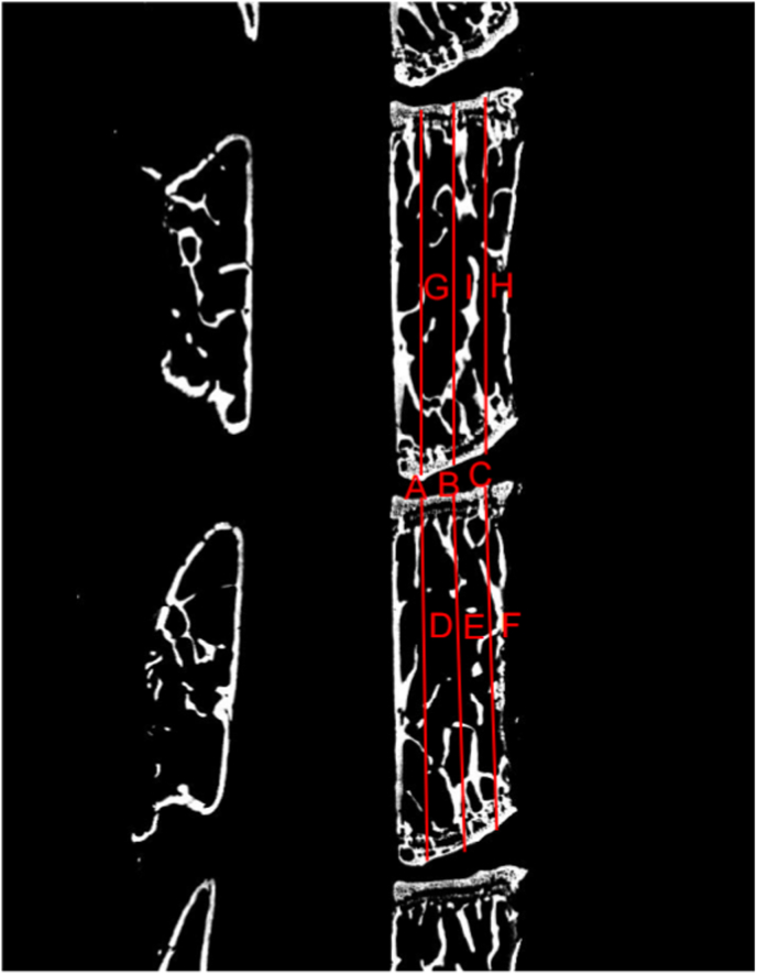
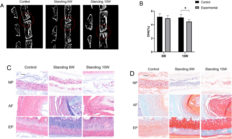
A standing mouse model was generated, and staining and imaging methods were used to evaluate the IDD model. Methylated RNA immunoprecipitation with next-generation sequencing (MeRIP-seq) was used to analyze mA methylation-associated transcripts in the NP, and real-time quantitative polymerase chain reaction (qRT-PCR) was used to detect the expression of methylation-related enzymes and conduct bio-informatics analysis.
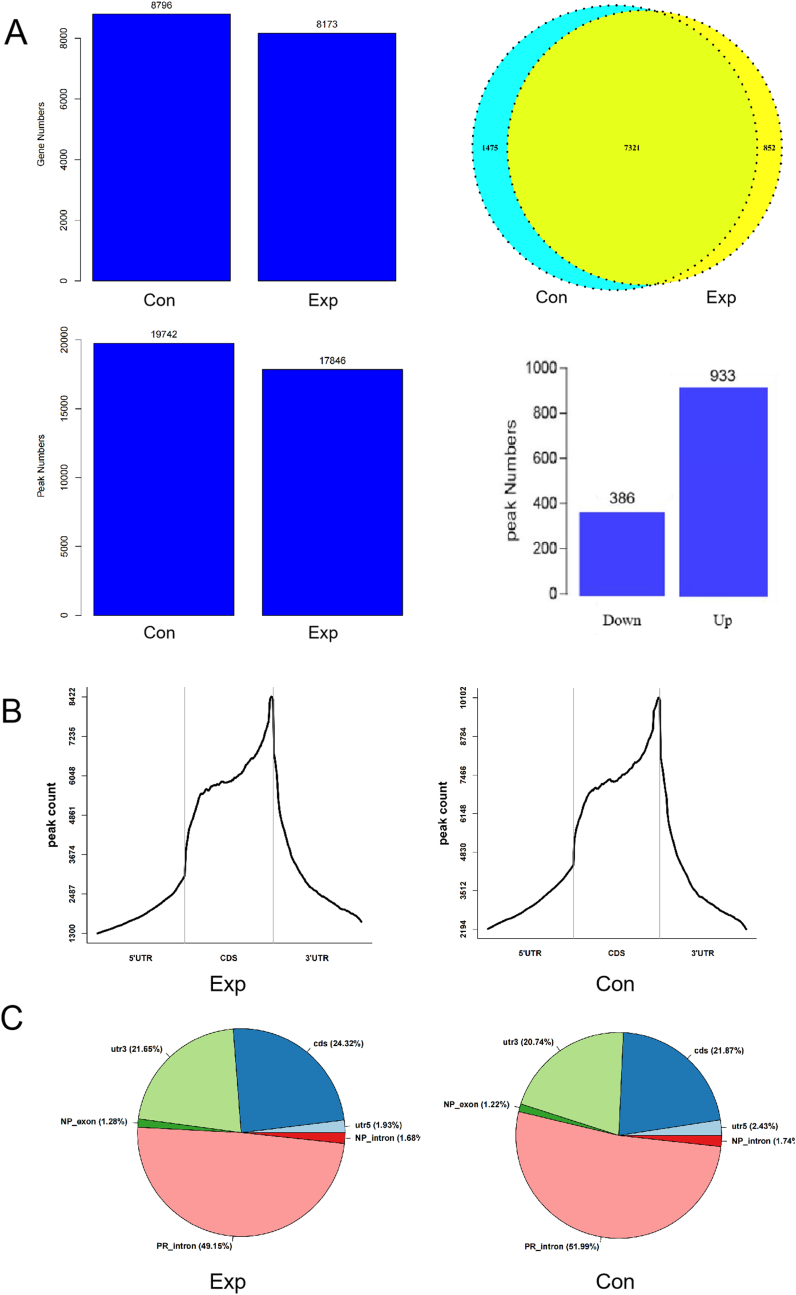
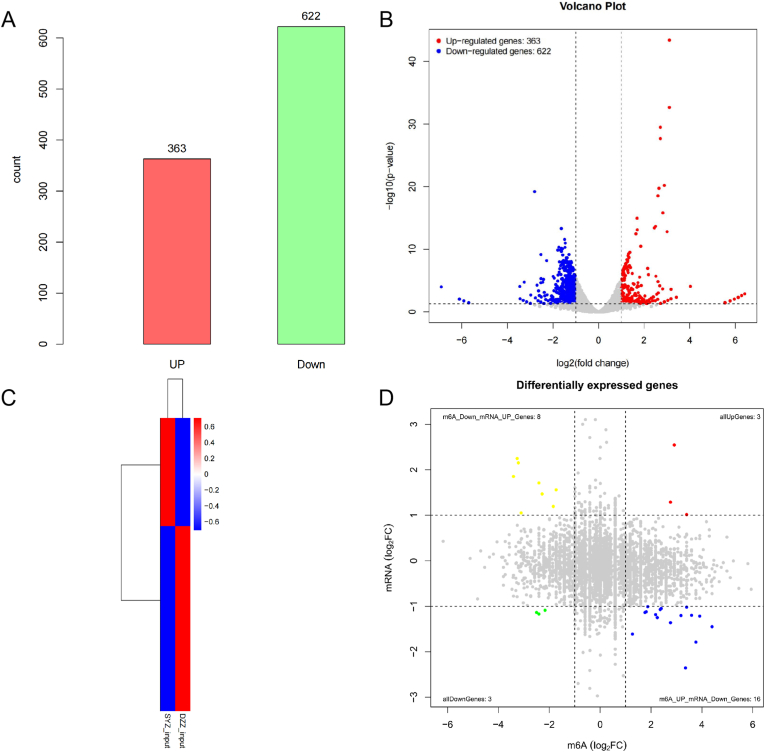
The standing mouse model caused IDD. Continuous axial pressure changed the expression of related methylases in degenerated NP tissue. Relative to the control group, the expression levels of KIAA1429, METTL14, METTL3, METTL4, WTAP, DGCR8, EIF3A and YTHDC1 in the experimental group were higher, while those of FTO, ELAVL1, HNRNPC1 and SRSF2 were lower. We identified 985 differentially expressed genes through MeRIP-Seq, among which 363 genes were significantly up-regulated, and 622 genes were significantly down-regulated. In addition, among the 9648 genes counted, 1319 mA peaks with significant differences in methylation were identified, among which 933 were significantly up-regulated, and 386 were significantly down-regulated. Genes and pathways that were enriched in IDD have been identified.
The results of this study elucidated the mA methylation pattern of NP tissue in degenerated lumbar intervertebral disc of mice and provided new perspectives and clues for research on and the treatment of lumbar disc degeneration.
As one of the important causes of low back and leg pain, intervertebral disc degeneration brings a huge economic burden to the society, family and medical system. Therefore, understanding the molecular and cellular mechanisms of intervertebral disc degeneration is of great significance for guiding clinical treatment. In this study, methylated RNA immunoprecipitation with next-generation sequencing on mice lumbar nucleus pulposus tissues found that differentially expressed genes and changes in the expression of related methylases, confirming that RNA methylation is involved in intervertebral disc degeneration. The process provides new vision and clues for future research on intervertebral disc degeneration.
研究椎间盘退变(IDD)过程中髓核(NP)组织的N6-甲基腺苷(m⁶A)修饰模式。
建立站立位小鼠模型,采用染色和成像方法评估IDD模型。利用甲基化RNA免疫沉淀结合下一代测序(MeRIP-seq)分析NP中与m⁶A甲基化相关的转录本,采用实时定量聚合酶链反应(qRT-PCR)检测甲基化相关酶的表达并进行生物信息学分析。
站立位小鼠模型导致了IDD。持续的轴向压力改变了退变NP组织中相关甲基化酶的表达。相对于对照组,实验组中KIAA1429、METTL14、METTL3、METTL4、WTAP、DGCR8、EIF3A和YTHDC1的表达水平较高,而FTO、ELAVL1、HNRNPC1和SRSF2的表达水平较低。通过MeRIP-Seq我们鉴定出985个差异表达基因,其中363个基因显著上调,622个基因显著下调。此外,在所统计的9648个基因中,鉴定出1319个甲基化存在显著差异的m⁶A峰,其中933个显著上调,386个显著下调。已确定在IDD中富集的基因和通路。
本研究结果阐明了小鼠退变腰椎椎间盘中NP组织的m⁶A甲基化模式,为腰椎间盘退变的研究和治疗提供了新的视角和线索。
作为腰腿痛的重要原因之一,椎间盘退变给社会、家庭和医疗系统带来了巨大的经济负担。因此,了解椎间盘退变的分子和细胞机制对指导临床治疗具有重要意义。在本研究中,对小鼠腰椎髓核组织进行甲基化RNA免疫沉淀结合下一代测序发现了差异表达基因以及相关甲基化酶表达的变化,证实RNA甲基化参与了椎间盘退变过程。该过程为未来椎间盘退变的研究提供了新的视野和线索。