• 文献检索
  • 文档翻译
  • 深度研究
  • 学术资讯
  • Suppr Zotero 插件Zotero 插件
  • 邀请有礼
  • 套餐&价格
  • 历史记录
应用&插件
Suppr Zotero 插件Zotero 插件浏览器插件Mac 客户端Windows 客户端微信小程序
定价
高级版会员购买积分包购买API积分包
服务
文献检索文档翻译深度研究API 文档MCP 服务
关于我们
关于 Suppr公司介绍联系我们用户协议隐私条款
关注我们

Suppr 超能文献

核心技术专利:CN118964589B侵权必究
粤ICP备2023148730 号-1Suppr @ 2026

文献检索

告别复杂PubMed语法,用中文像聊天一样搜索,搜遍4000万医学文献。AI智能推荐,让科研检索更轻松。

立即免费搜索

文件翻译

保留排版,准确专业,支持PDF/Word/PPT等文件格式,支持 12+语言互译。

免费翻译文档

深度研究

AI帮你快速写综述,25分钟生成高质量综述,智能提取关键信息,辅助科研写作。

立即免费体验

健康受试者黄斑区血管几何形态随性别和年龄的变化:一项眼底摄影研究。

Macular Vascular Geometry Changes With Sex and Age in Healthy Subjects: A Fundus Photography Study.

作者信息

Feng Ziqing, Wang Gengyuan, Xia Honghui, Li Meng, Liang Guoxia, Dong Tingting, Xiao Peng, Yuan Jin

机构信息

State Key Laboratory of Ophthalmology, Guangdong Provincial Key Laboratory of Ophthalmology and Visual Science, Guangdong Provincial Clinical Research Center for Ocular Diseases, Zhongshan Ophthalmic Center, Sun Yat-sen University, Guangzhou, China.

Department of Ophthalmology, Zhaoqing Gaoyao People's Hospital, Zhaoqing, China.

出版信息

Front Med (Lausanne). 2021 Dec 15;8:778346. doi: 10.3389/fmed.2021.778346. eCollection 2021.

DOI:10.3389/fmed.2021.778346
PMID:34977079
原文链接:https://pmc.ncbi.nlm.nih.gov/articles/PMC8714757/
Abstract

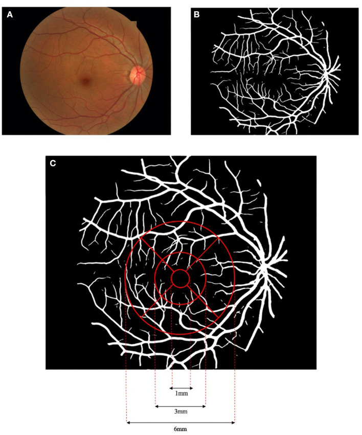
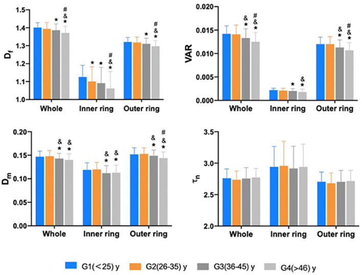
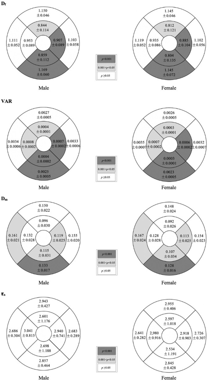
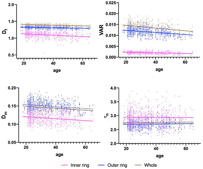
To characterize the sex- and age-related alterations of the macular vascular geometry in a population of healthy eyes using fundus photography. A cross-sectional study was conducted with 610 eyes from 305 healthy subjects (136 men, 169 women) who underwent fundus photography examination and was divided into four age groups (G1 with age ≤ 25 years, G2 with age 26-35 years, G3 with age 36-45 years, and G4 with age ≥ 46 years). A self-developed automated retinal vasculature analysis system allowed segmentation and separate multiparametric quantification of the macular vascular network according to the Early Treatment Diabetic Retinopathy Study (ETDRS). Vessel fractal dimension (D), vessel area rate (VAR), average vessel diameter (D), and vessel tortuosity (τ) were acquired and compared between sex and age groups. There was no significant difference between the mean age of male and female subjects (32.706 ± 10.372 and 33.494 ± 10.620, respectively, > 0.05) and the mean age of both sexes in each age group ( > 0.05). The D, VAR, and D of the inner ring, the D of the outer ring, and the D and VAR of the whole macula were significantly greater in men than women ( < 0.001, < 0.001, < 0.05, respectively). There was no significant change of τ between males and females ( > 0.05). The D, VAR, and D of the whole macula, the inner and outer rings associated negatively with age ( < 0.001), whereas the τ showed no significant association with age ( > 0.05). Comparison between age groups observed that D started to decrease from G2 compared with G1 in the inner ring ( < 0.05) and D, VAR, and D all decreased from G3 compared with the younger groups in the whole macula, inner and outer rings ( < 0.05). In the healthy subjects, macular vascular geometric parameters obtained from fundus photography showed that D, VAR, and D are related to sex and age while τ is not. The baseline values of the macular vascular geometry were also acquired for both sexes and all age groups.

摘要

利用眼底摄影技术,对健康人群中黄斑区血管几何形态的性别和年龄相关变化进行特征描述。对305名健康受试者(136名男性,169名女性)的610只眼睛进行了横断面研究,这些受试者均接受了眼底摄影检查,并被分为四个年龄组(G1组年龄≤25岁,G2组年龄26 - 35岁,G3组年龄36 - 45岁,G4组年龄≥46岁)。一个自行开发的自动化视网膜血管分析系统,能够根据早期糖尿病视网膜病变研究(ETDRS)对黄斑区血管网络进行分割和单独的多参数定量分析。获取血管分形维数(D)、血管面积率(VAR)、平均血管直径(D)和血管迂曲度(τ),并在性别和年龄组之间进行比较。男性和女性受试者的平均年龄之间无显著差异(分别为32.706±10.372和33.494±10.620,P>0.05),且各年龄组中两性的平均年龄之间也无显著差异(P>0.05)。男性内环的D、VAR和D、外环的D以及整个黄斑区的D和VAR均显著大于女性(分别为P<0.001、P<0.001、P<0.05)。男性和女性之间的τ无显著变化(P>0.05)。整个黄斑区的D、VAR和D以及内环和外环与年龄呈负相关(P<0.001),而τ与年龄无显著相关性(P>0.05)。年龄组之间的比较观察到,内环中与G1组相比,G2组的D开始降低(P<0.05),整个黄斑区、内环和外环中与较年轻组相比,G3组的D、VAR和D均降低(P<0.05)。在健康受试者中,通过眼底摄影获得的黄斑区血管几何参数表明,D、VAR和D与性别和年龄有关,而τ则无关。还获取了所有性别和年龄组的黄斑区血管几何形态的基线值。

https://cdn.ncbi.nlm.nih.gov/pmc/blobs/901e/8714757/f3a5643a44af/fmed-08-778346-g0004.jpg
https://cdn.ncbi.nlm.nih.gov/pmc/blobs/901e/8714757/fcc7cc156c0b/fmed-08-778346-g0001.jpg
https://cdn.ncbi.nlm.nih.gov/pmc/blobs/901e/8714757/8d5852c4acdc/fmed-08-778346-g0002.jpg
https://cdn.ncbi.nlm.nih.gov/pmc/blobs/901e/8714757/e9a3b0c614c9/fmed-08-778346-g0003.jpg
https://cdn.ncbi.nlm.nih.gov/pmc/blobs/901e/8714757/f3a5643a44af/fmed-08-778346-g0004.jpg

相似文献

1
Macular Vascular Geometry Changes With Sex and Age in Healthy Subjects: A Fundus Photography Study.健康受试者黄斑区血管几何形态随性别和年龄的变化:一项眼底摄影研究。
Front Med (Lausanne). 2021 Dec 15;8:778346. doi: 10.3389/fmed.2021.778346. eCollection 2021.
2
Retinal vascular geometry detection as a biomarker in diabetes mellitus.视网膜血管几何形态检测作为糖尿病的一种生物标志物
Eur J Ophthalmol. 2022 May;32(3):1710-1719. doi: 10.1177/11206721211033488. Epub 2021 Jul 20.
3
Combining functional and morphological retinal vascular characteristics achieves high-precision diagnosis of mild non-proliferative diabetic retinopathy.结合功能和形态视网膜血管特征可实现对轻度非增生性糖尿病视网膜病变的高精度诊断。
J Transl Med. 2024 Aug 28;22(1):798. doi: 10.1186/s12967-024-05597-7.
4
A spectrum of retinal vasculature measures and coronary artery disease.视网膜血管测量与冠状动脉疾病的关系。
Atherosclerosis. 2018 Jan;268:215-224. doi: 10.1016/j.atherosclerosis.2017.10.008. Epub 2017 Oct 10.
5
Optical coherence tomography angiography reveals progressive worsening of retinal vascular geometry in diabetic retinopathy and improved geometry after panretinal photocoagulation.光学相干断层扫描血管造影显示糖尿病视网膜病变的视网膜血管几何形状进行性恶化,全视网膜光凝治疗后几何形状改善。
PLoS One. 2019 Dec 30;14(12):e0226629. doi: 10.1371/journal.pone.0226629. eCollection 2019.
6
An extension of the Early Treatment Diabetic Retinopathy Study (ETDRS) system for grading of diabetic macular edema in the Astemizole Retinopathy Trial.阿咪唑视网膜病变试验中用于糖尿病性黄斑水肿分级的早期治疗糖尿病性视网膜病变研究(ETDRS)系统的扩展。
Curr Eye Res. 2006 Jun;31(6):535-47. doi: 10.1080/02713680600746112.
7
Comparison of the modified Early Treatment Diabetic Retinopathy Study and mild macular grid laser photocoagulation strategies for diabetic macular edema.改良早期糖尿病性视网膜病变研究与轻度黄斑格栅激光光凝术治疗糖尿病性黄斑水肿的策略比较
Arch Ophthalmol. 2007 Apr;125(4):469-80. doi: 10.1001/archopht.125.4.469.
8
Age-Related Alterations in Retinal Tissue Perfusion and Volumetric Vessel Density.年龄相关性视网膜组织灌注和容积血管密度的改变。
Invest Ophthalmol Vis Sci. 2019 Feb 1;60(2):685-693. doi: 10.1167/iovs.18-25864.
9
Comparison of Prevalence of Diabetic Macular Edema Based on Monocular Fundus Photography vs Optical Coherence Tomography.基于单眼眼底摄影与光学相干断层扫描技术的糖尿病性黄斑水肿患病率比较
JAMA Ophthalmol. 2016 Feb;134(2):222-8. doi: 10.1001/jamaophthalmol.2015.5332.
10
Reduction of retinal sensitivity in eyes with reticular pseudodrusen.网状假性小体相关的眼底敏感性降低。
Am J Ophthalmol. 2013 Dec;156(6):1184-1191.e2. doi: 10.1016/j.ajo.2013.06.036. Epub 2013 Aug 20.

引用本文的文献

1
Progress in clinical research and applications of retinal vessel quantification technology based on fundus imaging.基于眼底成像的视网膜血管量化技术的临床研究与应用进展
Front Bioeng Biotechnol. 2024 Feb 22;12:1329263. doi: 10.3389/fbioe.2024.1329263. eCollection 2024.
2
Understanding human aging and the fundamental cell signaling link in age-related diseases: the middle-aging hypovascularity hypoxia hypothesis.理解人类衰老以及与年龄相关疾病中的基本细胞信号传导联系:中年血管减少性缺氧假说。
Front Aging. 2023 Jun 13;4:1196648. doi: 10.3389/fragi.2023.1196648. eCollection 2023.

本文引用的文献

1
A novel multiple subdivision-based algorithm for quantitative assessment of retinal vascular tortuosity.一种新的基于多重细分的视网膜血管迂曲定量评估算法。
Exp Biol Med (Maywood). 2021 Oct;246(20):2222-2229. doi: 10.1177/15353702211032898. Epub 2021 Jul 25.
2
Retinal vascular geometry detection as a biomarker in diabetes mellitus.视网膜血管几何形态检测作为糖尿病的一种生物标志物
Eur J Ophthalmol. 2022 May;32(3):1710-1719. doi: 10.1177/11206721211033488. Epub 2021 Jul 20.
3
Quantitative analysis of inner retinal structural and microvascular alterations in intermediate age-related macular degeneration: A swept-source OCT angiography study.
定量分析中年相关性黄斑变性的内视网膜结构和微血管改变:扫频源 OCT 血管造影研究。
Photodiagnosis Photodyn Ther. 2020 Dec;32:102030. doi: 10.1016/j.pdpdt.2020.102030. Epub 2020 Oct 1.
4
Different Effect of Media Opacity on Vessel Density Measured by Different Optical Coherence Tomography Angiography Algorithms.不同光学相干断层扫描血管造影算法对血管密度测量的介质不透明度的影响不同。
Transl Vis Sci Technol. 2020 Jul 13;9(8):19. doi: 10.1167/tvst.9.8.19. eCollection 2020 Jul.
5
Retinal Vascular Tortuosity and Diameter Associations with Adiposity and Components of Body Composition.视网膜血管迂曲和直径与肥胖和身体成分的组成部分有关。
Obesity (Silver Spring). 2020 Sep;28(9):1750-1760. doi: 10.1002/oby.22885. Epub 2020 Jul 29.
6
Macular Functional and Morphological Changes in Intermediate Age-Related Maculopathy.中龄相关性黄斑病变的黄斑功能和形态变化。
Invest Ophthalmol Vis Sci. 2020 May 11;61(5):11. doi: 10.1167/iovs.61.5.11.
7
Inflammation mediates the relationship between obesity and retinal vascular calibre in 11-12 year-olds children and mid-life adults.炎症介导肥胖与 11-12 岁儿童和中年成年人视网膜血管口径之间的关系。
Sci Rep. 2020 Mar 19;10(1):5006. doi: 10.1038/s41598-020-61801-w.
8
Macular Vessel Density Measured With Optical Coherence Tomography Angiography and Its Associations in a Large Population-Based Study.利用光相干断层扫描血管造影术测量黄斑血管密度及其在大型人群研究中的相关性。
Invest Ophthalmol Vis Sci. 2019 Nov 1;60(14):4830-4837. doi: 10.1167/iovs.19-28137.
9
Normative database and determinants of macular vessel density measured by optical coherence tomography angiography.光学相干断层扫描血管造影测量的黄斑血管密度的规范数据库和决定因素。
Clin Exp Ophthalmol. 2020 Jan;48(1):44-52. doi: 10.1111/ceo.13648. Epub 2019 Oct 16.
10
Adverse reactions to fluorescein angiography: A comprehensive review of the literature.荧光素血管造影的不良反应:文献综述。
Surv Ophthalmol. 2019 Sep-Oct;64(5):679-693. doi: 10.1016/j.survophthal.2019.02.004. Epub 2019 Feb 14.