Dal-Bianco A, Schranzer R, Grabner G, Lanzinger M, Kolbrink S, Pusswald G, Altmann P, Ponleitner M, Weber M, Kornek B, Zebenholzer K, Schmied C, Berger T, Lassmann H, Trattnig S, Hametner S, Leutmezer F, Rommer P
Department of Neurology, Vienna, Austria.
Department of Medical Engineering, Carinthia University of Applied Sciences, Klagenfurt, Austria.
Front Neurol. 2021 Dec 21;12:632749. doi: 10.3389/fneur.2021.632749. eCollection 2021.
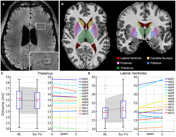
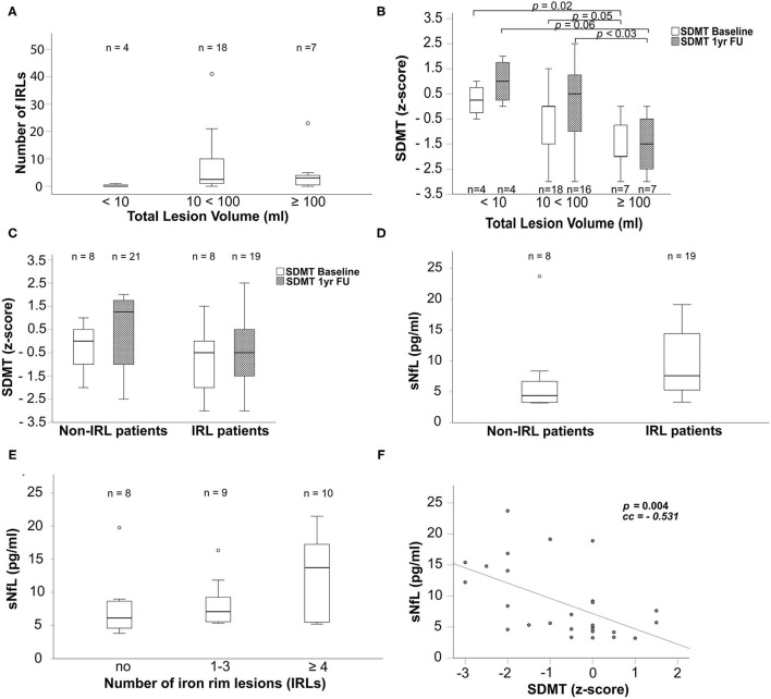
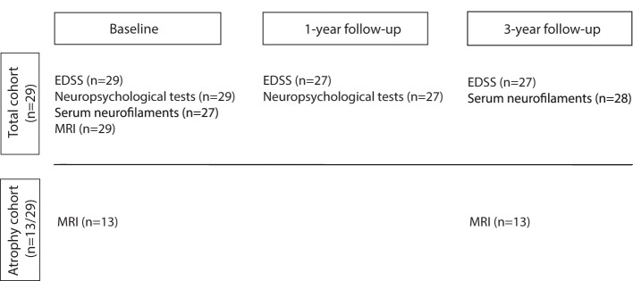
Multiple sclerosis (MS) is a demyelinating and neurodegenerative disease of the central nervous system, characterized by inflammatory-driven demyelination. Symptoms in MS manifest as both physical and neuropsychological deficits. With time, inflammation is accompanied by neurodegeneration, indicated by brain volume loss on an MRI. Here, we combined clinical, imaging, and serum biomarkers in patients with iron rim lesions (IRLs), which lead to severe tissue destruction and thus contribute to the accumulation of clinical disability. Subcortical atrophy and ventricular enlargement using an automatic segmentation pipeline for 7 Tesla (T) MRI, serum neurofilament light chain (sNfL) levels, and neuropsychological performance in patients with MS with IRLs and non-IRLs were assessed. In total 29 patients with MS [15 women, 24 relapsing-remitting multiple sclerosis (RRMS), and five secondary-progressive multiple sclerosis (SPMS)] aged 38 (22-69) years with an Expanded Disability Status Score of 2 (0-8) and a disease duration of 11 (5-40) years underwent neurological and neuropsychological examinations. Volumes of lesions, subcortical structures, and lateral ventricles on 7-T MRI (SWI, FLAIR, and MP2RAGE, 3D Segmentation Software) and sNfL concentrations using the Simoa SR-X Analyzer in IRL and non-IRL patients were assessed. (1) Iron rim lesions patients had a higher FLAIR lesion count ( = 0.047). Patients with higher MP2Rage lesion volume exhibited more IRLs ( <0.014) and showed poorer performance in the information processing speed tested within 1 year using the Symbol Digit Modalities Test (SDMT) ( <0.047). (2) Within 3 years, patients showed atrophy of the thalamus ( = 0.021) and putamen ( = 0.043) and enlargement of the lateral ventricles ( = 0.012). At baseline and after 3 years, thalamic volumes were lower in IRLs than in non-IRL patients ( = 0.045). (3) At baseline, IRL patients had higher sNfL concentrations ( = 0.028). Higher sNfL concentrations were associated with poorer SDMT ( = 0.004), regardless of IRL presence. (4) IRL and non-IRL patients showed no significant difference in the neuropsychological performance within 1 year. Compared with non-IRL patients, IRL patients had higher FLAIR lesion counts, smaller thalamic volumes, and higher sNfL concentrations. Our pilot study combines IRL and sNfL, two biomarkers considered indicative for neurodegenerative processes. Our preliminary data underscore the reported destructive nature of IRLs.
多发性硬化症(MS)是一种中枢神经系统的脱髓鞘和神经退行性疾病,其特征为炎症驱动的脱髓鞘。MS的症状表现为身体和神经心理缺陷。随着时间推移,炎症会伴随神经退行性变,这在MRI上表现为脑容量减少。在此,我们将临床、影像学和血清生物标志物相结合,研究患有铁环病变(IRL)的患者,这些病变会导致严重的组织破坏,从而促使临床残疾的累积。我们使用针对7特斯拉(T)MRI的自动分割流程评估了患有IRL和无IRL的MS患者的皮质下萎缩和脑室扩大情况、血清神经丝轻链(sNfL)水平以及神经心理表现。共有29例年龄为38(22 - 69)岁、扩展残疾状态评分2(0 - 8)且病程11(5 - 40)年的MS患者[15名女性,24例复发缓解型多发性硬化症(RRMS),5例继发进展型多发性硬化症(SPMS)]接受了神经学和神经心理学检查。使用7-T MRI(SWI、FLAIR和MP2RAGE,3D分割软件)评估了IRL和无IRL患者的病变、皮质下结构和侧脑室体积,并使用Simoa SR-X分析仪评估了sNfL浓度。(1)铁环病变患者的FLAIR病变计数更高(P = 0.047)。MP2Rage病变体积较大的患者表现出更多的IRL(P < 0.014),并且在使用符号数字模态测试(SDMT)在1年内测试的信息处理速度方面表现更差(P < 0.047)。(2)在3年内,患者出现丘脑萎缩(P = 0.021)和壳核萎缩(P = 0.043)以及侧脑室扩大(P = 0.012)。在基线和3年后,IRL患者的丘脑体积低于无IRL患者(P = 0.045)。(3)在基线时,IRL患者的sNfL浓度更高(P = 0.028)。无论是否存在IRL,较高的sNfL浓度都与较差的SDMT表现相关(P = 0.004)。(4)IRL和无IRL患者在1年内的神经心理表现无显著差异。与无IRL患者相比,IRL患者的FLAIR病变计数更高、丘脑体积更小且sNfL浓度更高。我们的初步研究将IRL和sNfL这两种被认为可指示神经退行性过程的生物标志物结合起来。我们的初步数据强调了IRL所报道的破坏性本质。