Hamad Shareef Suhayla, Abdel Aziz Ibrahim Ibrahim, Alzahrani Abdullah R, Al-Medhtiy Morteta H, Ameen Abdulla Mahmood
Department of Medical Microbiology, College of Science, Cihan University-Erbil, Erbil, Kurdistan Region, Iraq.
Department of Biology, College of Education, Salahaddin University-Erbil, Erbil, Kurdistan Region, Iraq.
Saudi J Biol Sci. 2022 Jan;29(1):564-573. doi: 10.1016/j.sjbs.2021.09.023. Epub 2021 Sep 16.
Since ancient times, herbal medicines have been applied in the treatment of cancer. Tea, derivative from the dried leaves of Camellia sinensis (L.) Kuntze plant is the most popular beverage globally after water and is available in various forms. Green tea has been expansively investigated for its beneficial properties of cancer prevention and therapy. The goal of the research: The current study was conducted to evaluate the hepaprotective character of methanolic green tea extract and its mechanism of action contrary to thioacetamide (TAA)-produced liver fibrosis of Sprague Dawley rats.
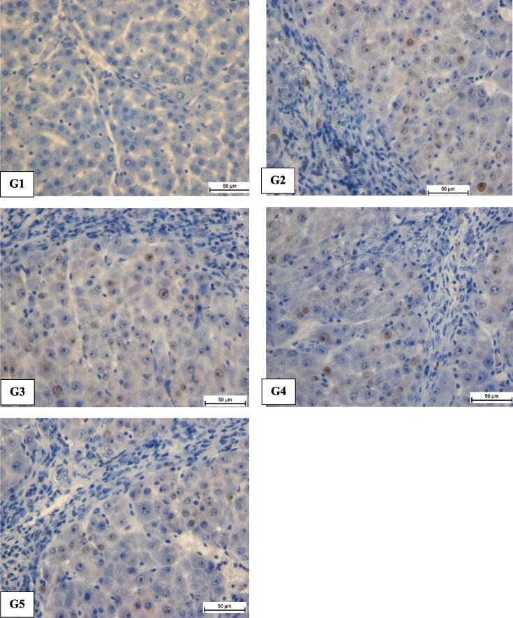
Thirty rodents were equally placed in 5 clusters including normal control, TAA group as a positive control, silymarin as standard drug control, and treatment groups consisting of high dose and a low dose Camellia sinensis. Rats in experimental clusters by mouth fed with C. sinensis at 250 mg/kg or 500 mg/kg daily for 2 months. After 60 days, all rats were sacrificed. Blood specimens were gathered for liver biochemical examination. Livers of all groups were dissected out and subjected to histopathological examination through the Hematoxylin and Eosin stain, Masson trichrome, and immunohistochemistry stains (PCNA). Liver tissue homogenate was also analyzed for antioxidant activity parameters.
Gross morphological examination showed a regular liver architecture in C. sinensis fed collections compared to the TAA sets. Histology of rat's liver fed with C. sinensis showed an important decrease in the liver index with hepatic cells propagation, mild cellular injury, and immunostaining showed significant down-expression of proliferating cell nuclear antigen (PCNA). TAA produced liver fibrosis through a significant increase in serum alanine transferase, aspartate aminotransferase, alkaline phosphatase, and bilirubin. Total protein and albumin also decreased in the TAA group. Moreover, the reduction of antioxidant enzyme activity including superoxide dismutase and catalase as well as the increase in malondialdehyde was detected in the TAA control group. Meanwhile, an abnormal level of liver biochemical parameters was restored closer to the normal levels in serum of the C. sinensis-fed clusters. In addition, C. sinensis fed assemblies showed elevated antioxidative enzymes activity with a reduction in malondialdehyde level comparable to the levels in silymarin-treated rats.
Green tea potentially inhibited the progression of liver cirrhosis, down -regulation of PCNA proliferation, prevented oxidation of hepatocytes, recovered SOD and CAT enzymes, condensed MDA and reduced cellular inflammation.
自古以来,草药就被用于癌症治疗。茶是由茶树(Camellia sinensis (L.) Kuntze)干燥叶子制成的衍生物,是全球仅次于水的最受欢迎饮品,有多种形式。绿茶因其预防和治疗癌症的有益特性而受到广泛研究。
本研究旨在评估甲醇绿茶提取物对硫代乙酰胺(TAA)诱导的Sprague Dawley大鼠肝纤维化的肝保护作用及其作用机制。
将30只啮齿动物平均分为5组,包括正常对照组、作为阳性对照的TAA组、作为标准药物对照的水飞蓟宾组以及由高剂量和低剂量茶树组成的治疗组。实验分组中的大鼠每天经口给予250mg/kg或500mg/kg的茶树,持续2个月。60天后,处死所有大鼠。采集血液样本进行肝脏生化检查。取出所有组的肝脏,通过苏木精和伊红染色、Masson三色染色和免疫组织化学染色(PCNA)进行组织病理学检查。还对肝组织匀浆进行抗氧化活性参数分析。
大体形态学检查显示,与TAA组相比,喂食茶树的组肝脏结构正常。喂食茶树的大鼠肝脏组织学检查显示肝脏指数显著降低,肝细胞增殖,轻度细胞损伤,免疫染色显示增殖细胞核抗原(PCNA)显著下调。TAA通过显著增加血清丙氨酸转氨酶、天冬氨酸转氨酶、碱性磷酸酶和胆红素导致肝纤维化。TAA组的总蛋白和白蛋白也降低。此外,在TAA对照组中检测到抗氧化酶活性降低,包括超氧化物歧化酶和过氧化氢酶,同时丙二醛增加。与此同时,喂食茶树的组血清中肝脏生化参数的异常水平恢复到更接近正常水平。此外,喂食茶树的组抗氧化酶活性升高,丙二醛水平降低,与水飞蓟宾治疗的大鼠水平相当。
绿茶可能抑制肝硬化的进展,下调PCNA增殖,防止肝细胞氧化,恢复SOD和CAT酶,浓缩MDA并减轻细胞炎症。