Gramuntell Yaiza, Klimczak Patrycja, Coviello Simona, Perez-Rando Marta, Nacher Juan
Neurobiology Unit, Program in Neurosciences and Institute of Biotechnology and Biomedicine (BIOTECMED), Universitat de València, Burjassot, Spain.
Spanish National Network for Research in Mental Health, Centro de Investigación Biomédica en Red de Salud Mental (CIBERSAM), Madrid, Spain.
Front Aging Neurosci. 2021 Dec 23;13:782737. doi: 10.3389/fnagi.2021.782737. eCollection 2021.
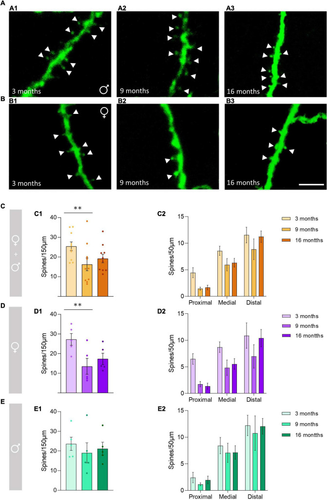
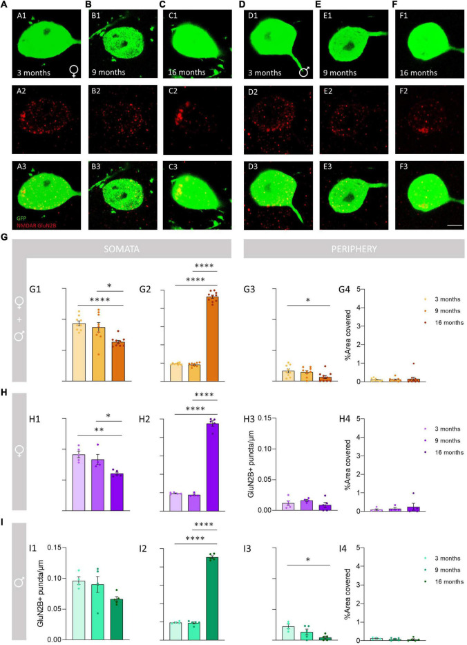
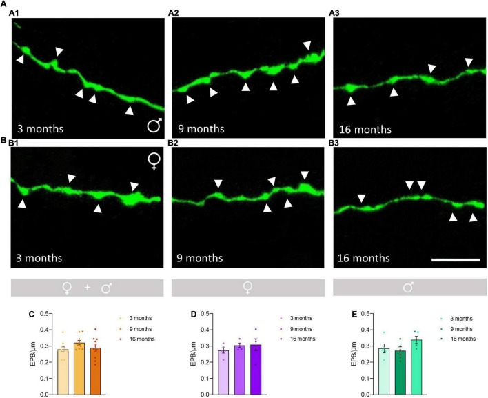
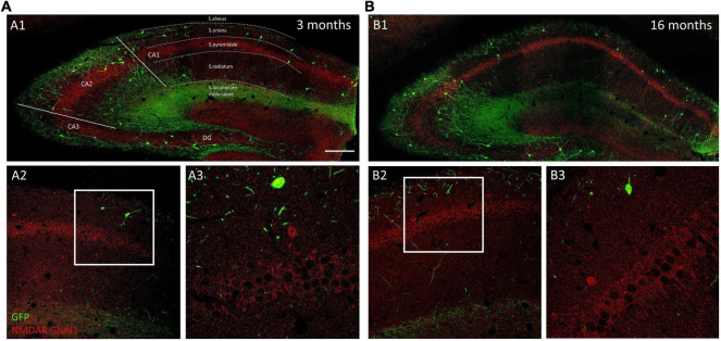
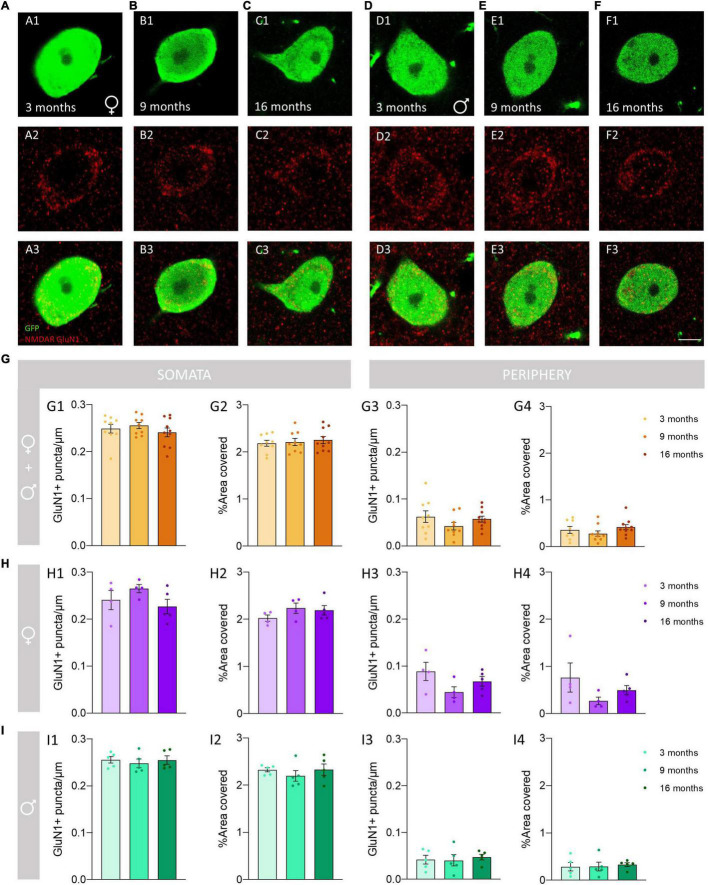
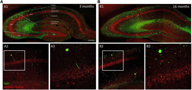
Changes in the physiology, neurochemistry and structure of neurons, particularly of their dendritic spines, are thought to be crucial players in age-related cognitive decline. One of the most studied brain structures affected by aging is the hippocampus, known to be involved in different essential cognitive processes. While the aging-associated quantitative changes in dendritic spines of hippocampal pyramidal cells have already been studied, the relationship between aging and the structural dynamics of hippocampal interneurons remains relatively unknown. Spines are not a frequent feature in cortical inhibitory neurons, but these postsynaptic structures are abundant in a subpopulation of somatostatin expressing interneurons, particularly in oriens-lacunosum moleculare (O-LM) cells in the hippocampal CA1. Previous studies from our laboratory have shown that the spines of these interneurons are highly plastic and influenced by NMDA receptor manipulation. Thus, in the present study, we have investigated the impact of aging on this interneuronal subpopulation. The analyses were performed in 3-, 9-, and 16-month-old GIN mice, a strain in which somatostatin positive interneurons express GFP. We studied the changes in the density of dendritic spines, , and the expression of NMDA receptors (GluN1 and GluN2B) using confocal microscopy and image analysis. We observed a significant decrease in dendritic spine density in 9-month-old animals when compared with 3-month-old animals. We also observed a decrease in the expression of the GluN2B subunit in O-LM cells, but not of that of GluN1, during aging. These results will constitute the basis for more advanced studies of the structure and connectivity of interneurons during aging and their contribution to cognitive decline.
神经元的生理、神经化学和结构变化,尤其是其树突棘的变化,被认为是与年龄相关的认知衰退的关键因素。受衰老影响研究最多的脑结构之一是海马体,已知其参与不同的重要认知过程。虽然已经对海马体锥体细胞树突棘的衰老相关定量变化进行了研究,但衰老与海马体中间神经元结构动力学之间的关系仍然相对未知。树突棘在皮质抑制性神经元中并不常见,但这些突触后结构在表达生长抑素的中间神经元亚群中丰富,特别是在海马体CA1区的分子层-腔隙层(O-LM)细胞中。我们实验室之前的研究表明,这些中间神经元的树突棘具有高度可塑性,并受NMDA受体操纵的影响。因此,在本研究中,我们研究了衰老对这个中间神经元亚群的影响。分析在3个月、9个月和16个月大的GIN小鼠中进行,该品系中生长抑素阳性中间神经元表达绿色荧光蛋白(GFP)。我们使用共聚焦显微镜和图像分析研究了树突棘密度的变化以及NMDA受体(GluN1和GluN2B)的表达。我们观察到,与3个月大的动物相比,9个月大的动物树突棘密度显著降低。我们还观察到,在衰老过程中,O-LM细胞中GluN2B亚基的表达下降,但GluN1的表达没有下降。这些结果将为更深入研究衰老过程中中间神经元的结构和连接性及其对认知衰退的贡献奠定基础。