• 文献检索
  • 文档翻译
  • 深度研究
  • 学术资讯
  • Suppr Zotero 插件Zotero 插件
  • 邀请有礼
  • 套餐&价格
  • 历史记录
应用&插件
Suppr Zotero 插件Zotero 插件浏览器插件Mac 客户端Windows 客户端微信小程序
定价
高级版会员购买积分包购买API积分包
服务
文献检索文档翻译深度研究API 文档MCP 服务
关于我们
关于 Suppr公司介绍联系我们用户协议隐私条款
关注我们

Suppr 超能文献

核心技术专利:CN118964589B侵权必究
粤ICP备2023148730 号-1Suppr @ 2026

文献检索

告别复杂PubMed语法,用中文像聊天一样搜索,搜遍4000万医学文献。AI智能推荐,让科研检索更轻松。

立即免费搜索

文件翻译

保留排版,准确专业,支持PDF/Word/PPT等文件格式,支持 12+语言互译。

免费翻译文档

深度研究

AI帮你快速写综述,25分钟生成高质量综述,智能提取关键信息,辅助科研写作。

立即免费体验

反转恢复预饱和零回波时间序列在软骨骨连接高对比成像中的可行性。

Feasibility of an Inversion Recovery-Prepared Fat-Saturated Zero Echo Time Sequence for High Contrast Imaging of the Osteochondral Junction.

机构信息

Department of Radiology, University of California, San Diego, CA, United States.

GE Healthcare, San Diego, CA, United States.

出版信息

Front Endocrinol (Lausanne). 2021 Dec 24;12:777080. doi: 10.3389/fendo.2021.777080. eCollection 2021.

DOI:10.3389/fendo.2021.777080
PMID:35002964
原文链接:https://pmc.ncbi.nlm.nih.gov/articles/PMC8739813/
Abstract

PURPOSE

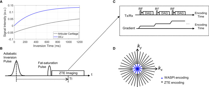
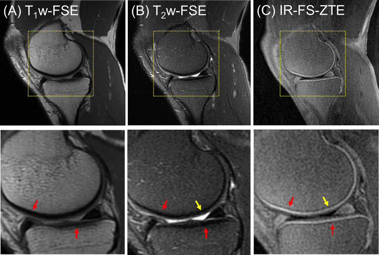
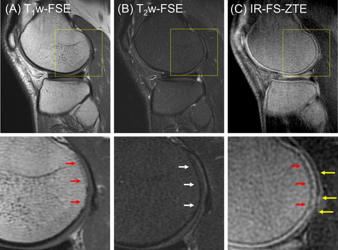
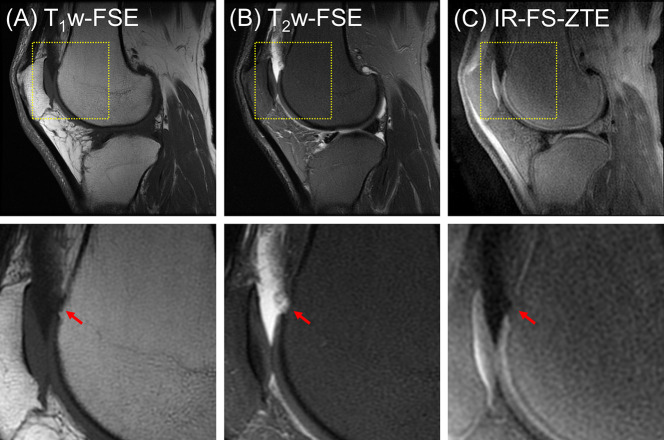
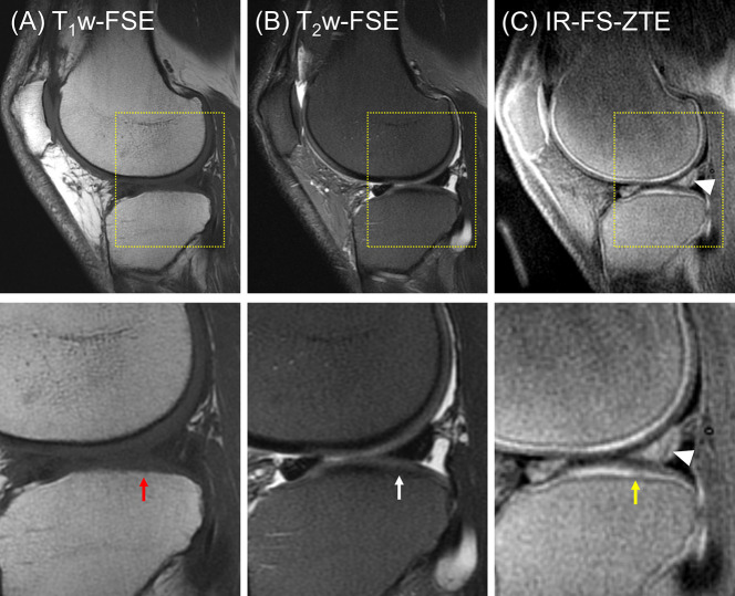
The osteochondral junction (OCJ) region-commonly defined to include the deep radial uncalcified cartilage, tidemark, calcified cartilage, and subchondral bone plate-functions to absorb mechanical stress and is commonly associated with the pathogenesis of osteoarthritis. However, magnetic resonance imaging of the OCJ region is difficult due to the tissues' short transverse relaxation times (i.e., short T or T*), which result in little or no signal with conventional MRI. The goal of this study is to develop a 3D adiabatic inversion recovery prepared fat saturated zero echo time (IR-FS-ZTE) sequence for high-contrast imaging of the OCJ.

METHOD

An IR-FS-ZTE MR sequence was developed to image the OCJ on a clinical 3T MRI scanner. The IR-FS-ZTE sequence employed an adiabatic inversion pulse followed by a fat saturation pulse that suppressed signals from the articular cartilage and fat. At an inversion time (TI) that was matched to the nulling point of the articular cartilage, continuous ZTE imaging was performed with a smoothly rotating readout gradient, which enabled time-efficient encoding of the OCJ region's short T signal with a minimal echo time (TE) of 12 μs. An ex vivo experiment with six cadaveric knee joints, and an experiment with six healthy volunteers and three patients with OA were performed to evaluate the feasibility of the proposed approach for high contrast imaging of the OCJ. Contrast-to-noise ratios (CNRs) between the OCJ and its neighboring femoral and tibial cartilage were measured.

RESULTS

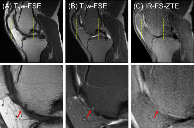
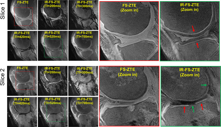
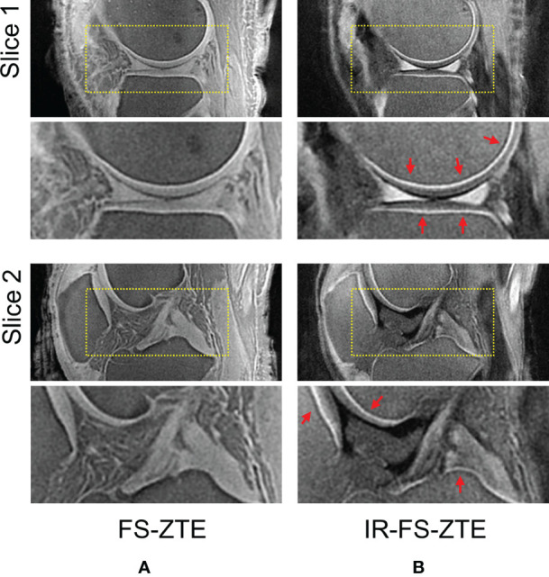
In the experiment, IR-FS-ZTE produced improved imaging of the OCJ region over the clinical sequences, and significantly improved the contrast compared to FS-ZTE without IR preparation (p = 0.0022 for tibial cartilage and p = 0.0019 for femoral cartilage with t-test). We also demonstrated the feasibility of high contrast imaging of the OCJ region using the proposed IR-FS-ZTE sequence, thereby providing more direct information on lesions in the OCJ. Clinical MRI did not detect signal from OCJ due to the long TE (>20 ms).

CONCLUSION

IR-FS-ZTE allows direct imaging of the OCJ region of the human knee and may help in elucidating the role of the OCJ in cartilage degeneration.

摘要

目的

骨软骨连接(OCJ)区域通常被定义为包括深层桡侧未钙化软骨、边界带、钙化软骨和软骨下骨板,其功能是吸收机械应力,通常与骨关节炎的发病机制有关。然而,由于 OCJ 区域的组织横向弛豫时间较短(即短 T 或 T*),常规 MRI 几乎没有或没有信号,因此对 OCJ 区域的磁共振成像(MRI)较为困难。本研究的目的是开发一种 3D 绝热反转恢复脂肪饱和零回波时间(IR-FS-ZTE)序列,以实现 OCJ 的高对比度成像。

方法

开发了一种 IR-FS-ZTE MR 序列,以在临床 3T MRI 扫描仪上对 OCJ 进行成像。IR-FS-ZTE 序列采用绝热反转脉冲,随后采用脂肪饱和脉冲,抑制关节软骨和脂肪的信号。在与关节软骨的零值点匹配的反转时间(TI)下,连续进行 ZTE 成像,采用平滑旋转的读出梯度,以 12μs 的最小回波时间(TE)对 OCJ 区域的短 T 信号进行高效编码。进行了一项包含 6 个尸体膝关节的离体实验和一项包含 6 个健康志愿者和 3 个 OA 患者的实验,以评估该方法用于 OCJ 高对比度成像的可行性。测量了 OCJ 与其相邻的股骨和胫骨软骨之间的对比噪声比(CNR)。

结果

在实验中,与临床序列相比,IR-FS-ZTE 对 OCJ 区域的成像效果得到了改善,并且与无 IR 准备的 FS-ZTE 相比,对比度显著提高(胫骨软骨 p=0.0022,股骨软骨 p=0.0019,t 检验)。我们还证明了使用所提出的 IR-FS-ZTE 序列对 OCJ 区域进行高对比度成像的可行性,从而提供了 OCJ 病变的更直接信息。由于较长的 TE(>20ms),临床 MRI 无法检测到 OCJ 的信号。

结论

IR-FS-ZTE 允许直接对人膝关节的 OCJ 区域进行成像,可能有助于阐明 OCJ 在软骨退变中的作用。

https://cdn.ncbi.nlm.nih.gov/pmc/blobs/30e1/8739813/6f2192eac889/fendo-12-777080-g008.jpg
https://cdn.ncbi.nlm.nih.gov/pmc/blobs/30e1/8739813/3dcbf3afef0f/fendo-12-777080-g001.jpg
https://cdn.ncbi.nlm.nih.gov/pmc/blobs/30e1/8739813/037215e6394c/fendo-12-777080-g002.jpg
https://cdn.ncbi.nlm.nih.gov/pmc/blobs/30e1/8739813/3cd20d8bc7a2/fendo-12-777080-g003.jpg
https://cdn.ncbi.nlm.nih.gov/pmc/blobs/30e1/8739813/34ace99895ef/fendo-12-777080-g004.jpg
https://cdn.ncbi.nlm.nih.gov/pmc/blobs/30e1/8739813/a6d18b9b289d/fendo-12-777080-g005.jpg
https://cdn.ncbi.nlm.nih.gov/pmc/blobs/30e1/8739813/dd55f6c95594/fendo-12-777080-g006.jpg
https://cdn.ncbi.nlm.nih.gov/pmc/blobs/30e1/8739813/44d7a376fe85/fendo-12-777080-g007.jpg
https://cdn.ncbi.nlm.nih.gov/pmc/blobs/30e1/8739813/6f2192eac889/fendo-12-777080-g008.jpg

相似文献

1
Feasibility of an Inversion Recovery-Prepared Fat-Saturated Zero Echo Time Sequence for High Contrast Imaging of the Osteochondral Junction.反转恢复预饱和零回波时间序列在软骨骨连接高对比成像中的可行性。
Front Endocrinol (Lausanne). 2021 Dec 24;12:777080. doi: 10.3389/fendo.2021.777080. eCollection 2021.
2
High-contrast osteochondral junction imaging using a 3D dual adiabatic inversion recovery-prepared ultrashort echo time cones sequence.采用三维双绝热反转恢复预备超短回波时间锥形序列进行高对比的软骨-骨连接成像。
NMR Biomed. 2021 Aug;34(8):e4559. doi: 10.1002/nbm.4559. Epub 2021 May 22.
3
Imaging of the region of the osteochondral junction (OCJ) using a 3D adiabatic inversion recovery prepared ultrashort echo time cones (3D IR-UTE-cones) sequence at 3 T.在 3T 磁共振成像系统中使用三维绝热反转恢复超短回波时间锥形(3D IR-UTE-cone)序列对骨软骨结合部(OCJ)进行成像。
NMR Biomed. 2019 May;32(5):e4080. doi: 10.1002/nbm.4080. Epub 2019 Feb 22.
4
Knee osteochondral junction imaging using a fast 3D T-weighted ultrashort echo time cones sequence at 3T.使用 3T 快速 3D T 加权超短回波时间锥形序列进行膝关节骨软骨连接成像。
Magn Reson Imaging. 2020 Nov;73:76-83. doi: 10.1016/j.mri.2020.08.003. Epub 2020 Aug 21.
5
Quantitative ultrashort echo time MR imaging of knee osteochondral junction: An ex vivo feasibility study.膝关节骨软骨结合部定量超短回波时间 MR 成像:一项离体可行性研究。
NMR Biomed. 2024 Dec;37(12):e5253. doi: 10.1002/nbm.5253. Epub 2024 Aug 28.
6
Inversion recovery zero echo time (IR-ZTE) imaging for direct myelin detection in human brain: a feasibility study.用于人脑直接髓鞘检测的反转恢复零回波时间(IR-ZTE)成像:一项可行性研究。
Quant Imaging Med Surg. 2020 May;10(5):895-906. doi: 10.21037/qims.2020.04.13.
7
High contrast cartilaginous endplate imaging using a 3D adiabatic inversion-recovery-prepared fat-saturated ultrashort echo time (3D IR-FS-UTE) sequence.采用三维绝热反转恢复预饱和脂肪抑制超短回波时间(3D IR-FS-UTE)序列进行高对比软骨终板成像。
NMR Biomed. 2021 Oct;34(10):e4579. doi: 10.1002/nbm.4579. Epub 2021 Jul 5.
8
High contrast cartilaginous endplate imaging in spine using three dimensional dual-inversion recovery prepared ultrashort echo time (3D DIR-UTE) sequence.使用三维双反转恢复预备超短回波时间(3D DIR-UTE)序列进行高对比度脊柱软骨终板成像。
Skeletal Radiol. 2024 May;53(5):881-890. doi: 10.1007/s00256-023-04503-4. Epub 2023 Nov 8.
9
3D-T prepared zero echo time-based PETRA sequence for in vivo biexponential relaxation mapping of semisolid short-T tissues at 3 T.3T 下用于半固态短 T 组织体内双指数弛豫测绘的 3D-T 准备零回波时间基于 PETRA 的序列。
J Magn Reson Imaging. 2019 Oct;50(4):1207-1218. doi: 10.1002/jmri.26664. Epub 2019 Jan 28.
10
IDEAL 3D spoiled gradient echo of the articular cartilage of the knee on 3.0 T MRI: a comparison with conventional 3.0 T fast spin-echo T2 fat saturation image.3.0T磁共振成像上膝关节软骨的理想三维扰相梯度回波成像:与传统3.0T快速自旋回波T2脂肪抑制图像的比较
Acta Radiol. 2015 Dec;56(12):1479-86. doi: 10.1177/0284185114556097. Epub 2014 Oct 27.

引用本文的文献

1
Comparison of pointwise encoding time reduction with radial acquisition (PETRA) imaging with conventional MR imaging for the diagnosis of traumatic fractures in children.用于儿童创伤性骨折诊断的逐点编码时间减少的径向采集(PETRA)成像与传统磁共振成像的比较。
BMC Med Imaging. 2025 Feb 18;25(1):55. doi: 10.1186/s12880-025-01594-4.
2
Detection of iron oxide nanoparticle (IONP)-labeled stem cells using quantitative ultrashort echo time imaging: a feasibility study.使用定量超短回波时间成像检测氧化铁纳米颗粒(IONP)标记的干细胞:一项可行性研究。
Quant Imaging Med Surg. 2023 Feb 1;13(2):585-597. doi: 10.21037/qims-22-654. Epub 2023 Jan 9.
3

本文引用的文献

1
Bright ultrashort echo time SWIFT MRI signal at the osteochondral junction is not located in the calcified cartilage.在骨软骨交界处,亮的超短回波时间 SWIFT MRI 信号并不位于钙化软骨中。
J Orthop Res. 2020 Dec;38(12):2649-2656. doi: 10.1002/jor.24777. Epub 2020 Jul 1.
2
Inversion recovery zero echo time (IR-ZTE) imaging for direct myelin detection in human brain: a feasibility study.用于人脑直接髓鞘检测的反转恢复零回波时间(IR-ZTE)成像:一项可行性研究。
Quant Imaging Med Surg. 2020 May;10(5):895-906. doi: 10.21037/qims.2020.04.13.
3
Whole-Brain Myelin Imaging Using 3D Double-Echo Sliding Inversion Recovery Ultrashort Echo Time (DESIRE UTE) MRI.
Making the invisible visible-ultrashort echo time magnetic resonance imaging: Technical developments and applications.
让不可见变为可见——超短回波时间磁共振成像:技术发展与应用
Appl Phys Rev. 2022 Dec;9(4):041303. doi: 10.1063/5.0086459.
4
Ultrashort echo time magnetic resonance imaging of the osteochondral junction.骨软骨连接的超短回波时间磁共振成像。
NMR Biomed. 2023 Feb;36(2):e4843. doi: 10.1002/nbm.4843. Epub 2022 Nov 20.
5
Articular Cartilage Assessment Using Ultrashort Echo Time MRI: A Review.关节软骨的超短回波时间 MRI 评估:综述。
Front Endocrinol (Lausanne). 2022 May 26;13:892961. doi: 10.3389/fendo.2022.892961. eCollection 2022.
使用 3D 双回波滑动反转恢复超短回波时间(DESIRE UTE)MRI 进行全脑髓鞘成像。
Radiology. 2020 Feb;294(2):362-374. doi: 10.1148/radiol.2019190911. Epub 2019 Nov 19.
4
Improved volumetric myelin imaging in human brain using 3D dual echo inversion recovery-prepared UTE with complex echo subtraction.使用具有复数回波减法的3D双回波反转恢复准备UTE改善人脑容积性髓鞘成像。
Magn Reson Med. 2020 Apr;83(4):1168-1177. doi: 10.1002/mrm.28082. Epub 2019 Nov 20.
5
Quantitative three-dimensional ultrashort echo time cones imaging of the knee joint with motion correction.膝关节运动校正的定量三维超短回波时间锥形成像。
NMR Biomed. 2020 Jan;33(1):e4214. doi: 10.1002/nbm.4214. Epub 2019 Nov 12.
6
Inversion recovery UTE based volumetric myelin imaging in human brain using interleaved hybrid encoding.基于反转恢复 UTE 的混合编码容积式人脑髓鞘成像。
Magn Reson Med. 2020 Mar;83(3):950-961. doi: 10.1002/mrm.27986. Epub 2019 Sep 18.
7
Imaging of the region of the osteochondral junction (OCJ) using a 3D adiabatic inversion recovery prepared ultrashort echo time cones (3D IR-UTE-cones) sequence at 3 T.在 3T 磁共振成像系统中使用三维绝热反转恢复超短回波时间锥形(3D IR-UTE-cone)序列对骨软骨结合部(OCJ)进行成像。
NMR Biomed. 2019 May;32(5):e4080. doi: 10.1002/nbm.4080. Epub 2019 Feb 22.
8
Towards quantification of myelin by solid-state MRI of the lipid matrix protons.通过脂质基质质子的固态 MRI 定量髓鞘。
Neuroimage. 2017 Dec;163:358-367. doi: 10.1016/j.neuroimage.2017.09.054. Epub 2017 Sep 28.
9
Short T imaging using a 3D double adiabatic inversion recovery prepared ultrashort echo time cones (3D DIR-UTE-Cones) sequence.使用三维双反转绝热恢复超短回波时间锥形(3D DIR-UTE-Cones)序列进行短 T 成像。
Magn Reson Med. 2018 May;79(5):2555-2563. doi: 10.1002/mrm.26908. Epub 2017 Sep 14.
10
Three-dimensional adiabatic inversion recovery prepared ultrashort echo time cones (3D IR-UTE-Cones) imaging of cortical bone in the hip.髋部皮质骨的三维绝热反转恢复准备的超短回波时间锥体(3D IR-UTE-Cones)成像。
Magn Reson Imaging. 2017 Dec;44:60-64. doi: 10.1016/j.mri.2017.07.012. Epub 2017 Jul 15.