Department of Radiology, Huadong Hospital, Fudan University, Shanghai, China.
Department of Otorhinolaryngology, Huadong Hospital, Fudan University, Shanghai, China.
Neural Plast. 2021 Dec 31;2021:2804533. doi: 10.1155/2021/2804533. eCollection 2021.
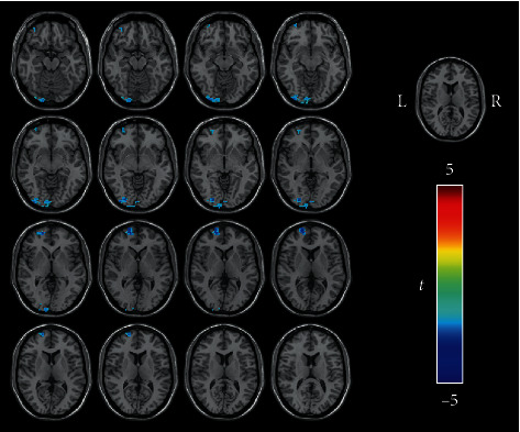
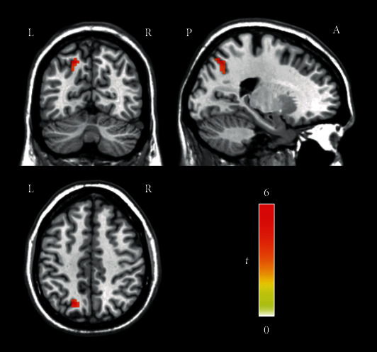
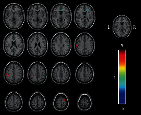
Previous functional magnetic resonance imaging (fMRI) analyses have shown that the dorsal attention network (DAN) is involved in the pathophysiological changes of tinnitus, but few relevant studies have been conducted, and the conclusions to date are not uniform. The purpose of this research was to test whether there is a change in intrinsic functional connectivity (FC) patterns between the DAN and other brain regions in tinnitus patients. Thirty-one patients with persistent tinnitus and thirty-three healthy controls were enrolled in this study. A group independent component analysis (ICA), degree centrality (DC) analysis, and seed-based FC analysis were conducted. In the group ICA, the tinnitus patients showed increased connectivity in the left superior parietal gyrus in the DAN compared to the healthy controls. Compared with the healthy controls, the tinnitus patients showed increased DC in the left inferior parietal gyrus and decreased DC in the left precuneus within the DAN. The clusters within the DAN with significant differences in the ICA or DC analysis between the tinnitus patients and the healthy controls were selected as regions of interest (ROIs) for seeds. The tinnitus patients exhibited significantly increased FC from the left superior parietal gyrus to several brain regions, including the left inferior parietal gyrus, the left superior marginal gyrus, and the right superior frontal gyrus, and decreased FC to the right anterior cingulate cortex. The tinnitus patients exhibited decreased FC from the left precuneus to the left inferior occipital gyrus, left calcarine cortex, and left superior frontal gyrus compared with the healthy controls. The findings of this study show that compared with healthy controls, tinnitus patients have altered functional connections not only within the DAN but also between the DAN and other brain regions. These results suggest that it may be possible to improve the disturbance and influence of tinnitus by regulating the DAN.
先前的功能磁共振成像(fMRI)分析表明,背侧注意网络(DAN)参与了耳鸣的病理生理变化,但相关研究较少,迄今为止的结论并不统一。本研究旨在测试耳鸣患者的 DAN 与其他脑区之间的内在功能连接(FC)模式是否发生变化。本研究纳入了 31 名持续性耳鸣患者和 33 名健康对照者。进行了组独立成分分析(ICA)、度中心性(DC)分析和种子点 FC 分析。在组 ICA 中,与健康对照组相比,耳鸣患者的 DAN 左侧顶上回的连接性增加。与健康对照组相比,耳鸣患者的 DAN 左侧顶下小叶的 DC 增加,左侧楔前叶的 DC 减少。在 ICA 或 DC 分析中,耳鸣患者与健康对照组之间存在显著差异的 DAN 内簇被选为种子的感兴趣区(ROI)。与健康对照组相比,耳鸣患者从左侧顶上回到包括左侧顶下小叶、左侧缘上回和右侧额上回在内的多个脑区的 FC 显著增加,而右侧前扣带皮层的 FC 减少。与健康对照组相比,耳鸣患者从左侧楔前叶到左侧枕下回、左侧距状回和左侧额上回的 FC 减少。本研究的结果表明,与健康对照组相比,耳鸣患者不仅在 DAN 内,而且在 DAN 与其他脑区之间的功能连接都发生了改变。这些结果表明,通过调节 DAN,可能改善耳鸣的干扰和影响。