• 文献检索
  • 文档翻译
  • 深度研究
  • 学术资讯
  • Suppr Zotero 插件Zotero 插件
  • 邀请有礼
  • 套餐&价格
  • 历史记录
应用&插件
Suppr Zotero 插件Zotero 插件浏览器插件Mac 客户端Windows 客户端微信小程序
定价
高级版会员购买积分包购买API积分包
服务
文献检索文档翻译深度研究API 文档MCP 服务
关于我们
关于 Suppr公司介绍联系我们用户协议隐私条款
关注我们

Suppr 超能文献

核心技术专利:CN118964589B侵权必究
粤ICP备2023148730 号-1Suppr @ 2026

文献检索

告别复杂PubMed语法,用中文像聊天一样搜索,搜遍4000万医学文献。AI智能推荐,让科研检索更轻松。

立即免费搜索

文件翻译

保留排版,准确专业,支持PDF/Word/PPT等文件格式,支持 12+语言互译。

免费翻译文档

深度研究

AI帮你快速写综述,25分钟生成高质量综述,智能提取关键信息,辅助科研写作。

立即免费体验

以腹水细胞学检查为金标准,不同计算机断层扫描征象鉴别恶性腹水与肝硬化腹水的诊断准确性

Diagnostic Accuracy of Different Computed Tomography Signs for Differentiating Between Malignant and Cirrhotic Ascites Keeping Ascitic Fluid Cytology as Gold Standard.

作者信息

Zafar Ibtesam, Majeed Ayesha Isani, Asad Muhammad Waseem, Khan Amir, Bhutta Muzammil Rasheed, Naeem Khan Muhammad Nasir

机构信息

Radiology, Pakistan Institute of Medical Sciences, Islamabad, PAK.

Gastroenterology and Hepatology, Pakistan Institute of Medical Sciences, Islamabad, PAK.

出版信息

Cureus. 2021 Dec 7;13(12):e20254. doi: 10.7759/cureus.20254. eCollection 2021 Dec.

DOI:10.7759/cureus.20254
PMID:35004063
原文链接:https://pmc.ncbi.nlm.nih.gov/articles/PMC8735839/
Abstract

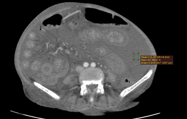
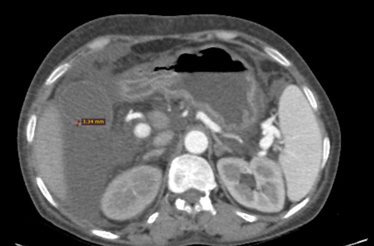
Objective The goal of this research was to define the diagnostic precision of CT signs to distinguish malignant ascites from cirrhotic ascites. Ascitic fluid cytology was kept as the gold standard. Study design This research was a prospective cross-sectional study. Place and duration of the study Participants' recruitment started on July 15, 2021, and the whole study lasted about three months till October 15, 2021, at the Radiology Department of Pakistan Institute of Medical Sciences, Islamabad. Patients and methods A total of 80 patients were included in the research and divided into two groups grounded on the cirrhotic or malignant etiology of the ascites based on their fluid cytology. Ascites volume, relative spread between the lesser sac and greater peritoneal cavity, the wall thickness of gallbladder, density of ascites, parietal peritoneum thickness and degree of its enhancement, and presence of septa and loculations were some of the major CT signs studied. Results The average age of patients included in this study was 36.2 ± 6.67 years (range 29-49 years). Of the 80 patients, 50 (62.5 %) were men, and 30 (37.5 %) were women. CT signs associated with the malignant ascites reported in this study were fluid present in the lesser sac (p = 0.03), peritoneal thickening and degree of its enhancement (p = 0.05), increased ascites density (p= 0.001), and presence of septa and loculations (63.6 % of malignant ascites). However, gallbladder wall thickness did not show any variation between both groups. Conclusion We conclude that in the diagnosis of malignant ascites, CT scan imaging can play a vital role. This research approves and testifies the benefits of indirect signs such as the spread of ascites, increased density of ascites, thickening and enhancement of parietal peritoneum, and ascitic fluid complexity in pointing out malignancy as a cause of ascites.

摘要

目的 本研究的目的是确定CT征象对鉴别恶性腹水和肝硬化腹水的诊断准确性。腹水细胞学检查作为金标准。

研究设计 本研究为前瞻性横断面研究。

研究地点和时间 研究对象招募于2021年7月15日开始,整个研究持续约三个月,至2021年10月15日结束,在伊斯兰堡巴基斯坦医学科学研究所放射科进行。

患者和方法 本研究共纳入80例患者,根据腹水的细胞学检查结果,基于肝硬化或恶性病因将其分为两组。研究的主要CT征象包括腹水容量、小网膜囊与大腹膜腔之间的相对扩散、胆囊壁厚度、腹水密度、壁层腹膜厚度及其强化程度,以及隔和分隔的存在情况。

结果 本研究纳入患者的平均年龄为36.2±6.67岁(范围29 - 49岁)。80例患者中,50例(62.5%)为男性,30例(37.5%)为女性。本研究报道的与恶性腹水相关的CT征象有小网膜囊内有液体(p = 0.03)、腹膜增厚及其强化程度(p = 0.05)、腹水密度增加(p = 0.001)以及隔和分隔的存在(恶性腹水中63.6%)。然而,两组之间胆囊壁厚度无差异。

结论 我们得出结论,在恶性腹水的诊断中,CT扫描成像可发挥重要作用。本研究证实并验证了间接征象如腹水扩散、腹水密度增加、壁层腹膜增厚和强化以及腹水复杂性在指出恶性肿瘤为腹水病因方面的益处。

https://cdn.ncbi.nlm.nih.gov/pmc/blobs/5a34/8735839/980e3e09d464/cureus-0013-00000020254-i02.jpg
https://cdn.ncbi.nlm.nih.gov/pmc/blobs/5a34/8735839/b3789eef14ce/cureus-0013-00000020254-i01.jpg
https://cdn.ncbi.nlm.nih.gov/pmc/blobs/5a34/8735839/980e3e09d464/cureus-0013-00000020254-i02.jpg
https://cdn.ncbi.nlm.nih.gov/pmc/blobs/5a34/8735839/b3789eef14ce/cureus-0013-00000020254-i01.jpg
https://cdn.ncbi.nlm.nih.gov/pmc/blobs/5a34/8735839/980e3e09d464/cureus-0013-00000020254-i02.jpg

相似文献

1
Diagnostic Accuracy of Different Computed Tomography Signs for Differentiating Between Malignant and Cirrhotic Ascites Keeping Ascitic Fluid Cytology as Gold Standard.以腹水细胞学检查为金标准,不同计算机断层扫描征象鉴别恶性腹水与肝硬化腹水的诊断准确性
Cureus. 2021 Dec 7;13(12):e20254. doi: 10.7759/cureus.20254. eCollection 2021 Dec.
2
Cirrhotic and malignant ascites: differential CT diagnosis.肝硬化和恶性腹水:CT 鉴别诊断。
Diagn Interv Imaging. 2012 May;93(5):365-70. doi: 10.1016/j.diii.2012.02.008. Epub 2012 Apr 27.
3
Diagnosing ascites: value of ascitic fluid total protein, albumin, cholesterol, their ratios, serum-ascites albumin and cholesterol gradient.腹水的诊断:腹水总蛋白、白蛋白、胆固醇及其比值、血清-腹水白蛋白梯度和血清-腹水胆固醇梯度的价值
J Gastroenterol Hepatol. 1995 May-Jun;10(3):295-9. doi: 10.1111/j.1440-1746.1995.tb01096.x.
4
Is MDCT an accurate tool to differentiate between benign and malignant etiology in diffuse peritoneal disease?MDCT 能否准确区分弥漫性腹膜疾病的良恶性病因?
Abdom Radiol (NY). 2022 Nov;47(11):3921-3929. doi: 10.1007/s00261-022-03641-y. Epub 2022 Aug 17.
5
[Concentration of lipids in ascitic fluid and the concentration gradient of albumin in blood and ascites: diagnostic significance].[腹水脂质浓度及血液与腹水白蛋白浓度梯度:诊断意义]
Ann Gastroenterol Hepatol (Paris). 1989 Nov;25(6):251-6.
6
Evaluation of fibronectin as a marker of malignant ascites.评估纤连蛋白作为恶性腹水标志物的作用。
J Gastroenterol Hepatol. 1992 Mar-Apr;7(2):161-4. doi: 10.1111/j.1440-1746.1992.tb00955.x.
7
Ascitic fluid analysis in malignancy-related ascites.恶性肿瘤相关性腹水的腹水分析
Hepatology. 1988 Sep-Oct;8(5):1104-9. doi: 10.1002/hep.1840080521.
8
Differentiation of benign from malignant induced ascites by measuring gallbladder wall thickness.通过测量胆囊壁厚度鉴别良性与恶性腹水。
Maedica (Bucur). 2011 Oct;6(4):282-6.
9
Biomarkers of malignant ascites-a myth or reality.恶性腹水的生物标志物——神话还是现实。
Med J Armed Forces India. 2011 Apr;67(2):108-12. doi: 10.1016/S0377-1237(11)60005-1. Epub 2011 Jul 21.
10
Usefulness of ascitic fluid cholesterol as a marker for malignant ascites.腹水胆固醇作为恶性腹水标志物的实用性。
Med Sci Monit. 2005 Mar;11(3):CR136-42.

引用本文的文献

1
Research progress and treatment status of malignant ascites.恶性腹水的研究进展与治疗现状
Front Oncol. 2024 Dec 16;14:1390426. doi: 10.3389/fonc.2024.1390426. eCollection 2024.
2
Possible use of 2D shear wave liver elastography in new-onset ascites evaluation.二维剪切波肝脏弹性成像在新发腹水评估中的可能应用。
BMC Gastroenterol. 2024 Feb 8;24(1):68. doi: 10.1186/s12876-024-03159-1.
3
The Evaluation Value of CT in the Efficacy of Neoadjuvant Chemotherapy in Ovarian Cancer Patients.CT 在卵巢癌患者新辅助化疗疗效评估中的价值。

本文引用的文献

1
The utility of texture-based classification of different types of ascites on magnetic resonance.基于纹理的磁共振不同类型腹水分类的效用。
J BUON. 2020 Mar-Apr;25(2):1237-1244.
2
Concave Margin Sign on CT as an Indication of Ascites.CT上的凹边征作为腹水的一种征象。
Radiol Case Rep. 2015 Nov 6;3(3):199. doi: 10.2484/rcr.v3i3.199. eCollection 2008.
3
CT imaging of peritoneal carcinomatosis and its mimics.腹膜癌病及其相似病症的CT成像
Contrast Media Mol Imaging. 2022 Jun 8;2022:7195888. doi: 10.1155/2022/7195888. eCollection 2022.
Diagn Interv Imaging. 2014 Sep;95(9):861-72. doi: 10.1016/j.diii.2014.02.009. Epub 2014 Mar 14.
4
Cirrhotic ascites review: Pathophysiology, diagnosis and management.肝硬化腹水综述:病理生理学、诊断与管理
World J Hepatol. 2013 May 27;5(5):251-63. doi: 10.4254/wjh.v5.i5.251.
5
Cirrhotic and malignant ascites: differential CT diagnosis.肝硬化和恶性腹水:CT 鉴别诊断。
Diagn Interv Imaging. 2012 May;93(5):365-70. doi: 10.1016/j.diii.2012.02.008. Epub 2012 Apr 27.
6
Omental cakes: unusual aetiologies and CT appearances.网膜饼:不寻常的病因及CT表现
Insights Imaging. 2011 Aug;2(4):399-408. doi: 10.1007/s13244-011-0105-4. Epub 2011 May 22.
7
Diagnosis and therapy of ascites in liver cirrhosis.肝硬化腹水的诊断与治疗。
World J Gastroenterol. 2011 Mar 14;17(10):1237-48. doi: 10.3748/wjg.v17.i10.1237.
8
The Role of 18F-FDG PET/CT in the evaluation of Ascites of Undetermined Origin.18F-FDG PET/CT在不明原因腹水评估中的作用
J Nucl Med. 2009 Apr;50(4):506-12. doi: 10.2967/jnumed.108.056382. Epub 2009 Mar 16.
9
Detection of peritoneal metastases.腹膜转移的检测
Cancer Imaging. 2001 Feb 1;1(2):4-12. doi: 10.1102/1470-7330.2001.002.
10
The role of ultrasonography and computed tomography in determining the etiology of ascites.超声检查和计算机断层扫描在确定腹水病因中的作用。
Saudi Med J. 2007 Dec;28(12):1822-6.