Department of Chemistry, Massachusetts Institute of Technology, 170 Albany Street, Cambridge, Massachusetts 02139, United States.
Department of Chemistry and Biochemistry, Wichita State University, 1845 Fairmount St., Wichita, Kansas 67260, United States.
J Am Chem Soc. 2022 Jan 26;144(3):1416-1430. doi: 10.1021/jacs.1c12056. Epub 2022 Jan 11.
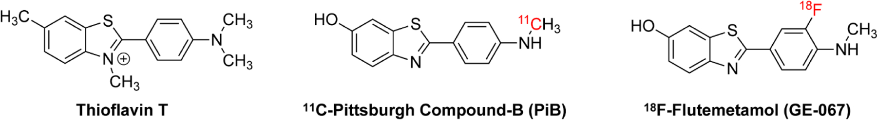
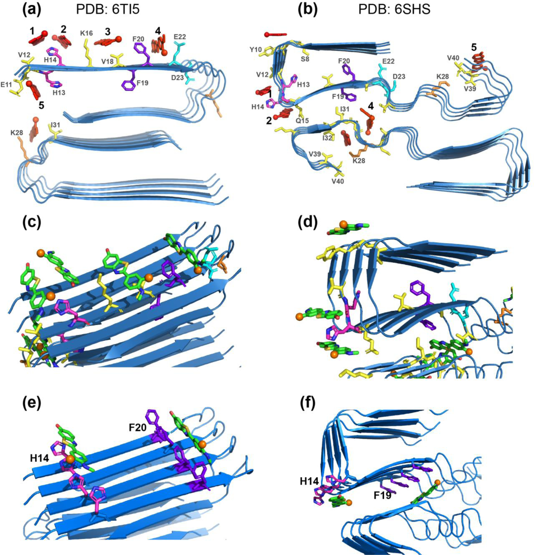
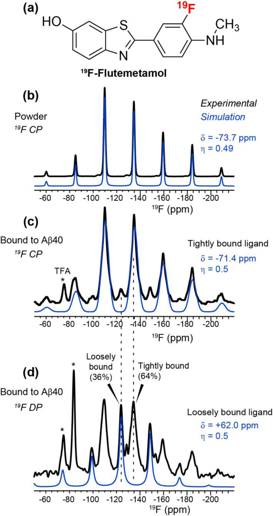
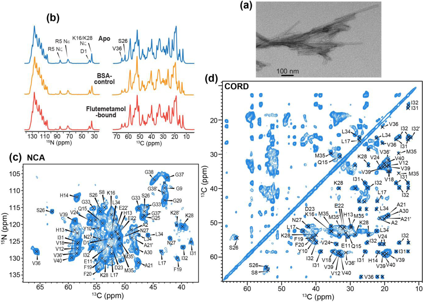
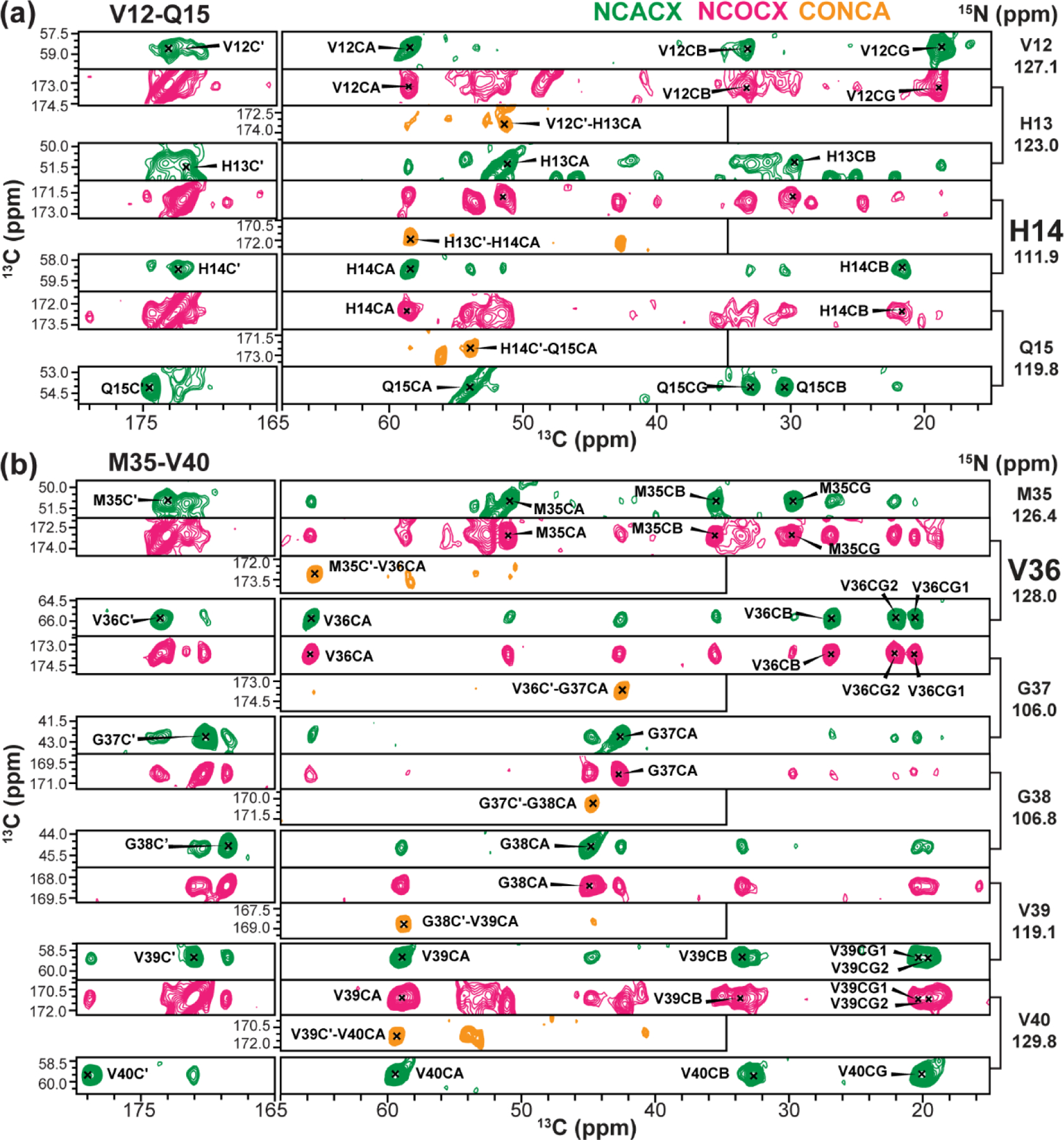
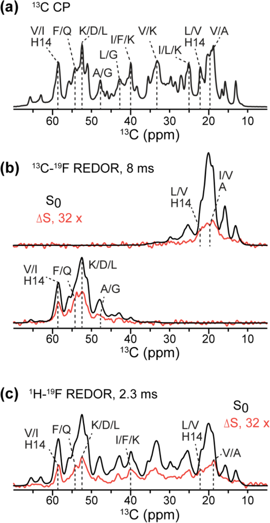
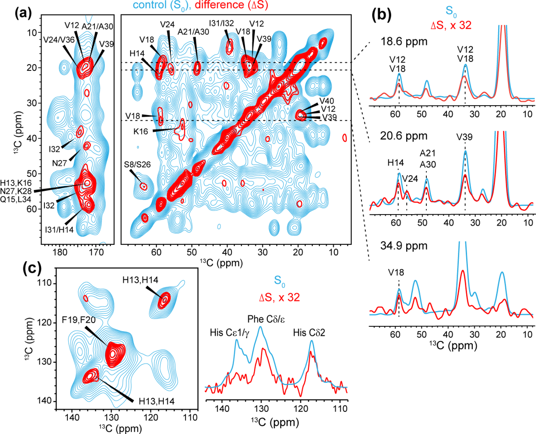
Amyloid imaging by positron emission tomography (PET) is an important method for diagnosing neurodegenerative disorders such as Alzheimer's disease. Many C- and F-labeled PET tracers show varying binding capacities, specificities, and affinities for their target proteins. The structural basis of these variations is poorly understood. Here we employ F and C solid-state NMR to investigate the binding sites of a PET ligand, flutemetamol, to the 40-residue Alzheimer's β-amyloid peptide (Aβ40). Analytical high-performance liquid chromatography and F NMR spectra show that flutemetamol binds the current Aβ40 fibril polymorph with a stoichiometry of one ligand per four to five peptides. Half of the ligands are tightly bound while the other half are loosely bound. C and N chemical shifts indicate that this Aβ40 polymorph has an immobilized N-terminus, a non-β-sheet His14, and a non-β-sheet C-terminus. We measured the proximity of the ligand fluorine to peptide residues using F-C and F-H rotational-echo double-resonance (REDOR) experiments. The spectra show that three segments in the peptide, VHH, VFF, and VV, lie the closest to the ligand. REDOR-constrained docking simulations indicate that these three segments form multiple binding sites, and the ligand orientations and positions at these sites are similar across different Aβ polymorphs. Comparison of the flutemetamol-interacting residues in Aβ40 with the small-molecule binding sites in other amyloid proteins suggest that conjugated aromatic compounds preferentially bind β-sheet surface grooves lined by aromatic, polar, and charged residues. These motifs may explain the specificity of different PET tracers to different amyloid proteins.
正电子发射断层扫描(PET)的淀粉样蛋白成像是诊断神经退行性疾病(如阿尔茨海默病)的重要方法。许多 C 和 F 标记的 PET 示踪剂对其靶蛋白表现出不同的结合能力、特异性和亲和力。这些变化的结构基础知之甚少。在这里,我们使用 F 和 C 固态 NMR 研究 PET 配体 flutemetamol 与 40 个残基的阿尔茨海默氏β-淀粉样肽(Aβ40)的结合位点。分析高效液相色谱和 F NMR 谱表明,flutemetamol 以一个配体与四到五个肽结合的比例与当前的 Aβ40 原纤维多晶型结合。一半的配体紧密结合,另一半则松散结合。C 和 N 化学位移表明,这种 Aβ40 多晶型具有固定的 N 末端、非β-折叠 His14 和非β-折叠 C 末端。我们使用 F-C 和 F-H 旋转回波双共振(REDOR)实验测量了配体氟与肽残基的接近程度。谱表明,肽中的三个片段 VHH、VFF 和 VV 与配体最接近。REDOR 约束对接模拟表明,这三个片段形成了多个结合位点,并且这些位点的配体取向和位置在不同的 Aβ 多晶型之间相似。与其他淀粉样蛋白中的小分子结合位点相比,flutemetamol 相互作用的残基在 Aβ40 中的位置表明,共轭芳族化合物优先与由芳族、极性和带电残基组成的β-折叠表面凹槽结合。这些基序可以解释不同 PET 示踪剂对不同淀粉样蛋白的特异性。