Department of Radiology, University of Florida, College of Medicine, Gainesville, FL, 32610, USA.
Geriatrician (PP), Silivrikapi Mh. Hisaralti Cd, Istanbul, 34093, Turkey.
Nat Commun. 2022 Jan 11;13(1):203. doi: 10.1038/s41467-021-27887-0.
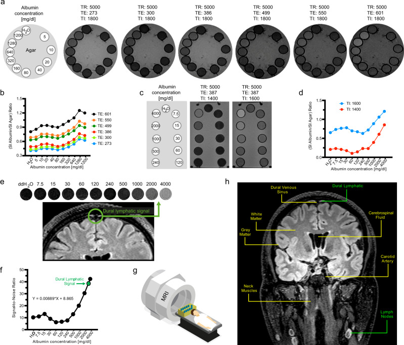
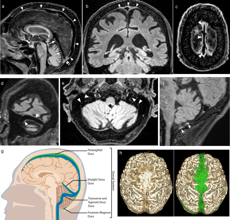
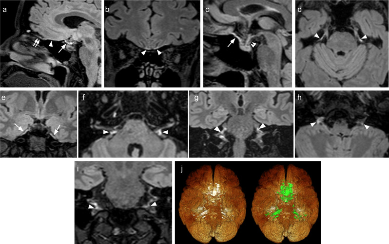
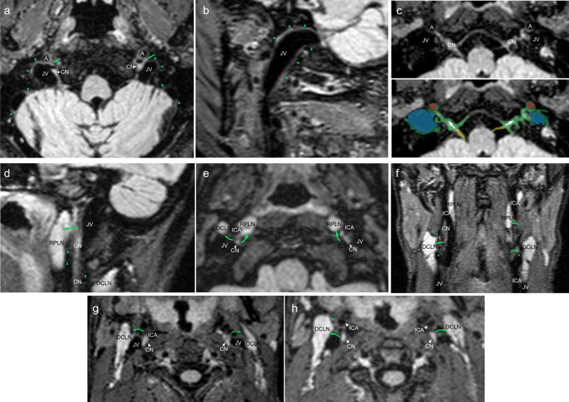
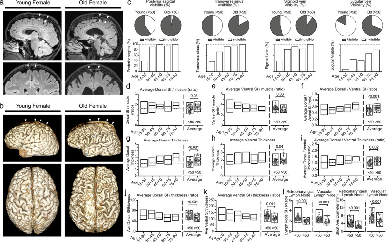
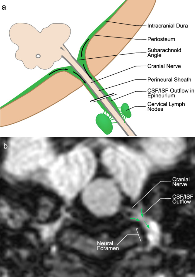
Meningeal lymphatic vessels have been described in animal studies, but limited comparable data is available in human studies. Here we show dural lymphatic structures along the dural venous sinuses in dorsal regions and along cranial nerves in the ventral regions in the human brain. 3D T2-Fluid Attenuated Inversion Recovery magnetic resonance imaging relies on internal signals of protein rich lymphatic fluid rather than contrast media and is used in the present study to visualize the major human dural lymphatic structures. Moreover we detect direct connections between lymphatic fluid channels along the cranial nerves and vascular structures and the cervical lymph nodes. We also identify age-related cervical lymph node atrophy and thickening of lymphatics channels in both dorsal and ventral regions, findings which reflect the reduced lymphatic output of the aged brain.
脑膜淋巴管在动物研究中已有描述,但在人类研究中可用的可比数据有限。在这里,我们在人脑的背侧区域沿硬脑膜静脉窦以及在腹侧区域沿颅神经显示硬脑膜淋巴管结构。3D T2- 液体衰减反转恢复磁共振成像是依靠富含蛋白质的淋巴液的内部信号,而不是对比剂,本研究中用于可视化主要的人类硬脑膜淋巴管结构。此外,我们还检测到沿颅神经的淋巴液通道与血管结构和颈部淋巴结之间的直接连接。我们还发现与年龄相关的颈部淋巴结萎缩和背侧和腹侧区域的淋巴管通道增厚,这些发现反映了老年大脑的淋巴输出减少。