Zheng Hui, Zhang Hanfei, Wang Shan, Xiao Feng, Liao Meiyan
Zhongnan Hospital, Wuhan University, Wuhan, China.
Front Genet. 2022 Jan 6;12:783391. doi: 10.3389/fgene.2021.783391. eCollection 2021.
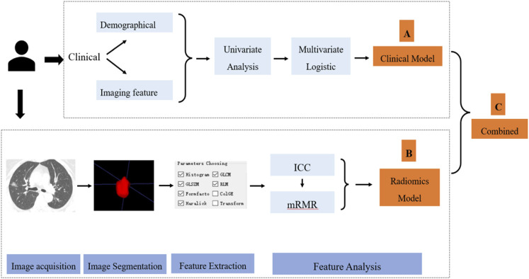
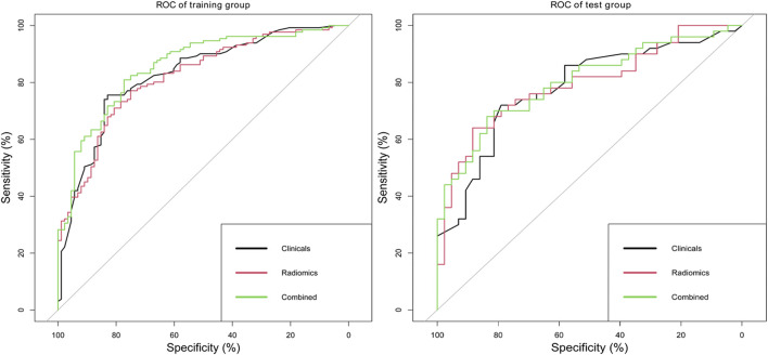
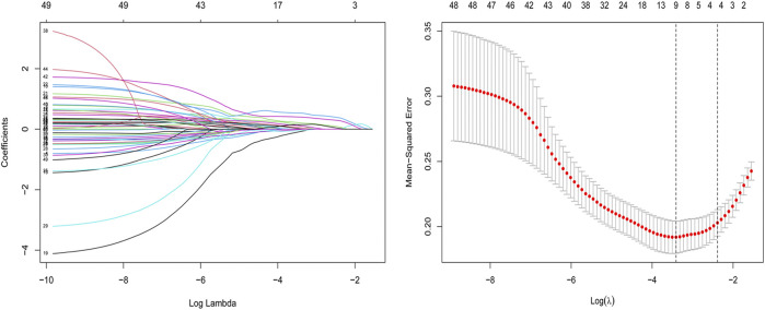
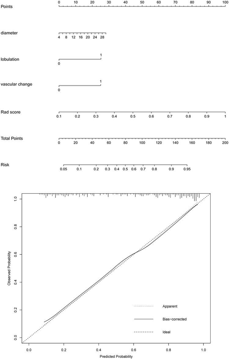
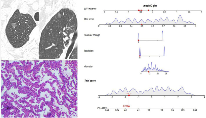
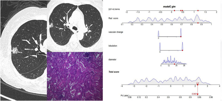
To explore the diagnostic value of CT radiographic images and radiomics features for invasive classification of lung adenocarcinoma manifesting as ground-glass nodules (GGNs) in computer tomography (CT). A total of 312 GGNs were enrolled in this retrospective study. All GGNs were randomly divided into training set ( = 219) and test set ( = 93). Univariate and multivariate logistic regressions were used to establish a clinical model, while the minimum redundancy maximum relevance (mRMR) and least absolute shrinkage and selection operator (LASSO) algorithm were used to select the radiomics features and construct the radiomics model. A combined model was finally built by combining these two models. The performance of these models was assessed in both training and test set. A combined nomogram was developed based on the combined model and evaluated with its calibration curves and C-index. Diameter [odds ratio (OR), 1.159; < 0.001], lobulation (OR, 2.953; = 0.002), and vascular changes (OR, 3.431; < 0.001) were retained as independent predictors of the invasive adenocarcinoma (IAC) group. Eleven radiomics features were selected by mRMR and LASSO method to established radiomics model. The clinical model and radiomics mode showed good predictive ability in both training set and test set. When two models were combined, the diagnostic area under the curve (AUC) value was higher than the single clinical or radiomics model (training set: 0.86 vs. 0.83 vs. 0.82; test set: 0.80 vs. 0.78 vs. 0.79). The constructed combined nomogram could effectively quantify the risk degree of 3 image features and Rad score with a C-index of 0.855 (95%: 0.805∼0.905). Radiographic and radiomics features show high accuracy in the invasive diagnosis of GGNs, and their combined analysis can improve the diagnostic efficacy of IAC manifesting as GGNs. The nomogram, serving as a noninvasive and accurate predictive tool, can help judge the invasiveness of GGNs prior to surgery and assist clinicians in creating personalized treatment strategies.
探讨计算机断层扫描(CT)中CT影像学图像和影像组学特征对表现为磨玻璃结节(GGN)的肺腺癌侵袭性分类的诊断价值。本回顾性研究共纳入312个GGN。所有GGN被随机分为训练集(n = 219)和测试集(n = 93)。采用单因素和多因素逻辑回归建立临床模型,同时采用最小冗余最大相关性(mRMR)和最小绝对收缩和选择算子(LASSO)算法选择影像组学特征并构建影像组学模型。最终通过将这两个模型相结合构建联合模型。在训练集和测试集中评估这些模型的性能。基于联合模型开发联合列线图,并通过其校准曲线和C指数进行评估。直径[比值比(OR),1.159;P<0.001]、分叶(OR,2.953;P = 0.002)和血管改变(OR,3.431;P<0.001)被保留为浸润性腺癌(IAC)组的独立预测因子。通过mRMR和LASSO方法选择11个影像组学特征以建立影像组学模型。临床模型和影像组学模型在训练集和测试集中均显示出良好的预测能力。当两个模型相结合时,曲线下面积(AUC)值高于单一临床或影像组学模型(训练集:0.86对0.83对0.82;测试集:0.80对0.78对0.79)。构建的联合列线图能够有效量化3个影像特征和Rad评分的风险程度,C指数为0.855(95%:0.805~0.905)。影像学和影像组学特征在GGN的侵袭性诊断中显示出较高的准确性,它们的联合分析可以提高表现为GGN的IAC的诊断效能。列线图作为一种无创且准确的预测工具,可在手术前帮助判断GGN的侵袭性,并协助临床医生制定个性化治疗策略。