Science for Life Laboratory, Department of Medical Biochemistry and Microbiology, Uppsala Universitygrid.8993.b, Uppsala, Sweden.
Department of Cell and Molecular Biology, Uppsala Universitygrid.8993.b, Uppsala, Sweden.
mBio. 2021 Feb 22;13(1):e0002222. doi: 10.1128/mbio.00022-22. Epub 2022 Feb 1.
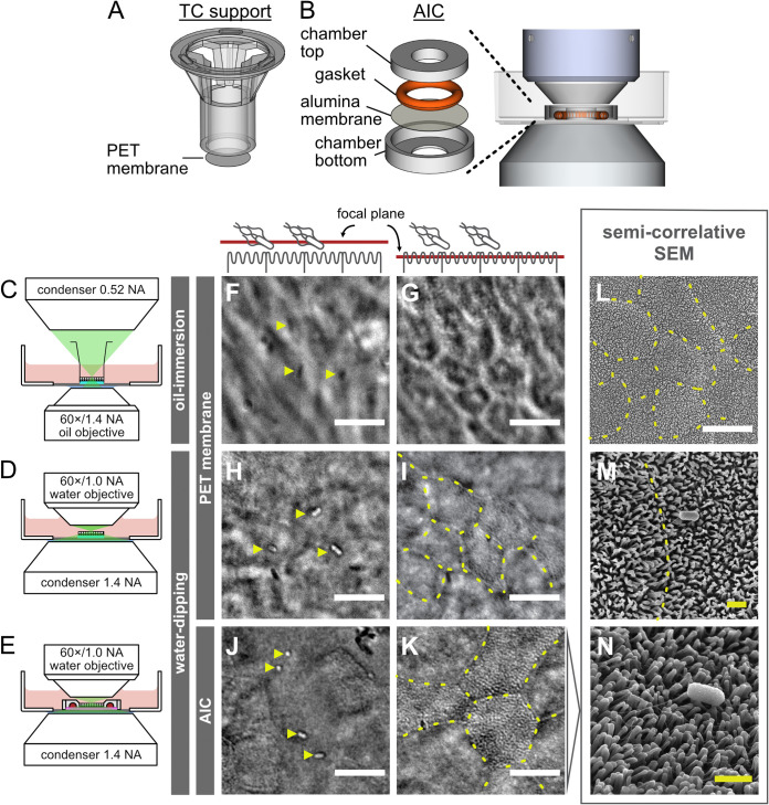
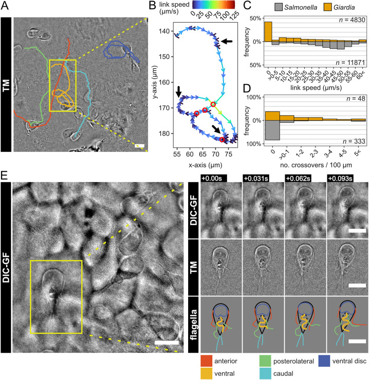
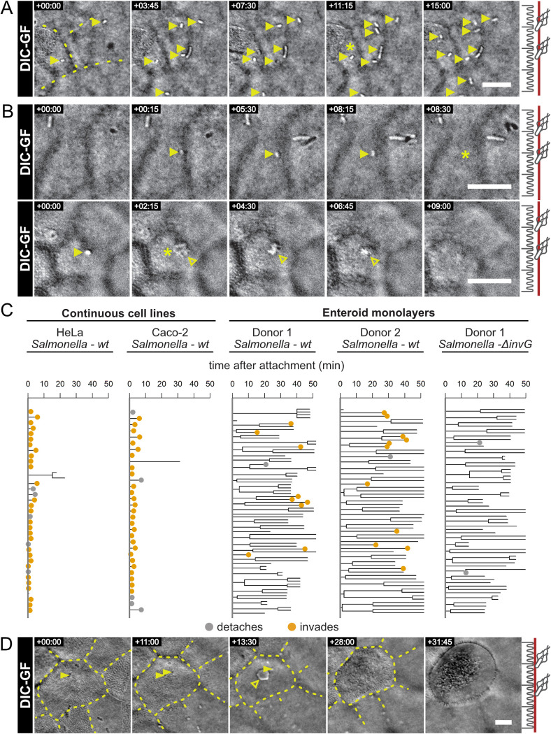
Interactions between individual pathogenic microbes and host tissues involve fast and dynamic processes that ultimately impact the outcome of infection. Using live-cell microscopy, these dynamics can be visualized to study, e.g., microbe motility, binding and invasion of host cells, and intrahost-cell survival. Such methodology typically employs confocal imaging of fluorescent tags in tumor-derived cell line infections on glass. This allows high-definition imaging but poorly reflects the host tissue's physiological architecture and may result in artifacts. We developed a method for live-cell imaging of microbial infection dynamics on human adult stem cell-derived intestinal epithelial cell (IEC) layers. These IEC layers are grown in apical imaging chambers, optimized for physiological cell arrangement and fast, but gentle, differential interference contrast (DIC) imaging. This allows subsecond visualization of both microbial and epithelial surface ultrastructure at high resolution without using fluorescent reporters. We employed this technology to probe the behavior of two model pathogens, Salmonella enterica serovar Typhimurium and Giardia intestinalis, at the intestinal epithelial surface. Our results reveal pathogen-specific swimming patterns on the epithelium and show that Salmonella lingers on the IEC surface for prolonged periods before host cell invasion, while Giardia uses circular swimming with intermittent attachments to scout for stable adhesion sites. The method even permits tracking of individual Giardia flagella, demonstrating that active flagellar beating and attachment to the IEC surface are not mutually exclusive. This work describes a generalizable and relatively inexpensive approach to resolving dynamic pathogen-IEC layer interactions, applicable even to genetically nontractable microorganisms. Knowledge of dynamic niche-specific interactions between single microbes and host cells is essential to understand infectious disease progression. However, advances in this field have been hampered by the inherent conflict between the technical requirements for high-resolution live-cell imaging on the one hand and conditions that best mimic physiological infection niche parameters on the other. Toward bridging this divide, we present a methodology for differential interference contrast (DIC) imaging of pathogen interactions at the apical surface of enteroid-derived intestinal epithelia, providing both high spatial and temporal resolution. This alleviates the need for fluorescent reporters in live-cell imaging and provides dynamic information about microbe interactions with a nontransformed, confluent, polarized, and microvilliated human gut epithelium. Using this methodology, we uncover previously unrecognized stages of Salmonella and Giardia infection cycles at the epithelial surface.
个体病原微生物与宿主组织之间的相互作用涉及快速和动态的过程,这些过程最终会影响感染的结果。使用活细胞显微镜,可以对这些动态过程进行可视化观察,例如研究微生物的运动、与宿主细胞的结合和入侵以及细胞内生存。这种方法通常使用肿瘤衍生细胞系感染玻璃上的荧光标记物进行共聚焦成像。这允许进行高清晰度成像,但不能很好地反映宿主组织的生理结构,并且可能导致伪影。我们开发了一种用于在人成年干细胞衍生的肠上皮细胞 (IEC) 层上进行微生物感染动力学的活细胞成像方法。这些 IEC 层在顶侧成像室中生长,该成像室经过优化,可实现生理细胞排列和快速但温和的微分干涉对比 (DIC) 成像。这允许在不使用荧光报告器的情况下,以高分辨率对微生物和上皮表面超微结构进行亚秒级可视化。我们利用这项技术来探测两种模型病原体——沙门氏菌血清型 Typhimurium 和肠道贾第虫——在肠上皮表面的行为。我们的结果揭示了病原体在肠上皮表面的特定游动模式,并表明沙门氏菌在宿主细胞入侵之前会在 IEC 表面长时间停留,而贾第虫则使用圆形游动并间歇性附着来寻找稳定的附着位点。该方法甚至允许跟踪单个贾第虫鞭毛,证明活跃的鞭毛跳动和与 IEC 表面的附着并不相互排斥。这项工作描述了一种可推广且相对廉价的方法,可以解决动态病原体-IEC 层相互作用的问题,即使对于遗传上难以处理的微生物也适用。了解单个微生物和宿主细胞之间动态生态位特异性相互作用对于理解传染病的进展至关重要。然而,该领域的进展受到高分辨率活细胞成像的技术要求与最佳模拟生理感染生态位参数的条件之间固有冲突的阻碍。为了弥合这一鸿沟,我们提出了一种用于肠类器官衍生的肠上皮细胞顶表面上病原体相互作用的微分干涉对比 (DIC) 成像方法,提供了高空间和时间分辨率。这减轻了活细胞成像中对荧光报告器的需求,并提供了关于微生物与非转化、连续、极化和微绒毛化的人类肠道上皮相互作用的动态信息。使用这种方法,我们在肠上皮表面发现了以前未被识别的沙门氏菌和贾第虫感染周期阶段。