Department of Cardiology, Zhongda Hospital, Southeast University, Hunan Road, Nanjing, 210009 Jiangsu, China.
Oxid Med Cell Longev. 2022 Jan 25;2022:2785113. doi: 10.1155/2022/2785113. eCollection 2022.
Myocardial ischemia/reperfusion (I/R) injury can aggravate myocardial injury. Programmed necrosis plays a crucial role in this injury. However, the role of exosomal miRNAs in myocardial I/R injury remains unclear. Therefore, this study is aimed at exploring the function and mechanism of exosomal miR-17-3p in myocardial I/R injury.
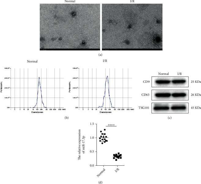
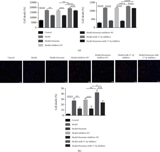
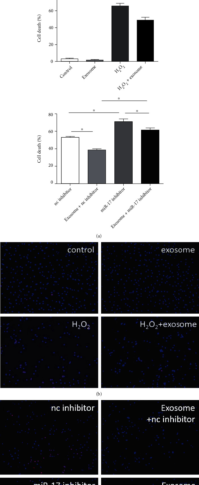
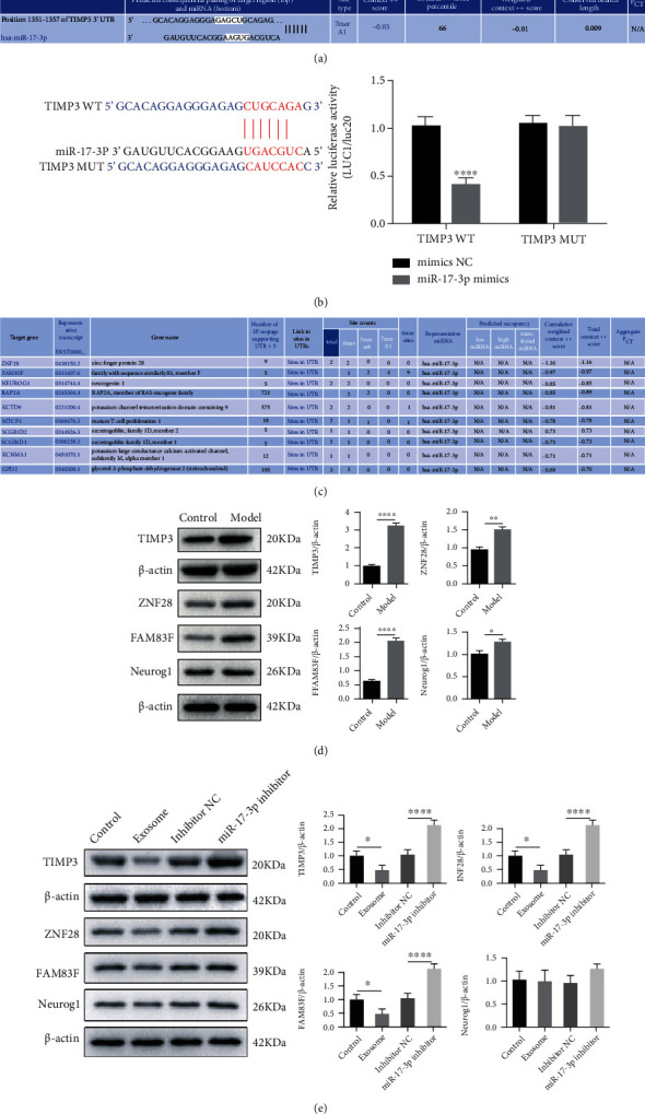
The myocardial I/R injury animal model was established in C57BL/6 mice. Exosomes were identified using transmission electron microscopy (TEM), nanoparticle tracking analysis (NTA), and Western blotting. Programmed necrosis was detected by PI staining. Heart function and myocardial infarct size were evaluated using echocardiography and triphenyl tetrazolium chloride (TTC) staining, respectively. Histopathological changes were visualized by hematoxylin and eosin (H&E) and Masson staining. The regulation of TIMP3 expression by miR-17-3p was verified using a dual-luciferase reporter assay. Lactate dehydrogenase (LDH) and tumor necrosis factor- (TNF-) levels were measured by enzyme-linked immunosorbent assays (ELISA). TIMP3 expression was measured by quantitative reverse transcription-polymerase chain reaction (qRT-PCR) and Western blotting.
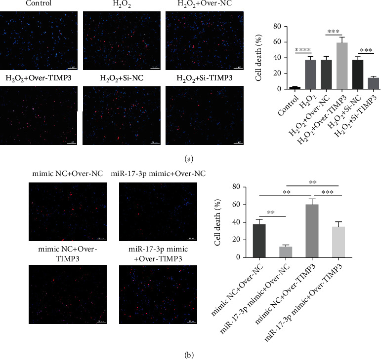
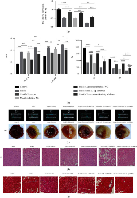
We demonstrated that miR-17-3p was significantly downregulated in peripheral blood exosomes after cardiac I/R injury. Further analysis indicated that exosomal miR-17-3p attenuated HO-induced programmed necrosis in cardiomyocytes in vitro. Moreover, TIMP3 was a target for miR-17-3p. TIMP3 affected HO-induced programmed necrosis in cardiomyocytes. This effect was modulated by miR-17-3p in vitro. Furthermore, exosomal miR-17-3p greatly alleviated cardiac I/R injury in vivo.
The present study demonstrated that exosomal miR-17-3p alleviated the programmed necrosis associated with cardiac I/R injury by regulating TIMP3 expression. These findings could represent a potential treatment for I/R injury.
心肌缺血/再灌注(I/R)损伤可加重心肌损伤。程序性细胞坏死在这种损伤中起着关键作用。然而,外泌体 miRNAs 在心肌 I/R 损伤中的作用尚不清楚。因此,本研究旨在探讨外泌体 miR-17-3p 在心肌 I/R 损伤中的功能和机制。
在 C57BL/6 小鼠中建立心肌 I/R 损伤动物模型。使用透射电子显微镜(TEM)、纳米颗粒跟踪分析(NTA)和 Western blot 鉴定外泌体。通过碘化丙啶(PI)染色检测程序性细胞坏死。通过超声心动图和三苯基四氮唑(TTC)染色评估心功能和心肌梗死面积。通过苏木精和伊红(H&E)和马松染色观察组织病理学变化。通过双荧光素酶报告实验验证 miR-17-3p 对 TIMP3 表达的调节。通过酶联免疫吸附测定(ELISA)测量乳酸脱氢酶(LDH)和肿瘤坏死因子-(TNF-)水平。通过定量逆转录聚合酶链反应(qRT-PCR)和 Western blot 测量 TIMP3 表达。
我们表明,心脏 I/R 损伤后外周血外泌体中的 miR-17-3p 显著下调。进一步分析表明,外泌体 miR-17-3p 可减轻体外心肌细胞中 HO 诱导的程序性细胞坏死。此外,TIMP3 是 miR-17-3p 的靶标。TIMP3 影响心肌细胞中 HO 诱导的程序性细胞坏死。这种作用可通过 miR-17-3p 在体外进行调节。此外,外泌体 miR-17-3p 可显著减轻体内心脏 I/R 损伤。
本研究表明,外泌体 miR-17-3p 通过调节 TIMP3 表达减轻与心脏 I/R 损伤相关的程序性细胞坏死。这些发现可为 I/R 损伤的治疗提供新的靶点。