• 文献检索
  • 文档翻译
  • 深度研究
  • 学术资讯
  • Suppr Zotero 插件Zotero 插件
  • 邀请有礼
  • 套餐&价格
  • 历史记录
应用&插件
Suppr Zotero 插件Zotero 插件浏览器插件Mac 客户端Windows 客户端微信小程序
定价
高级版会员购买积分包购买API积分包
服务
文献检索文档翻译深度研究API 文档MCP 服务
关于我们
关于 Suppr公司介绍联系我们用户协议隐私条款
关注我们

Suppr 超能文献

核心技术专利:CN118964589B侵权必究
粤ICP备2023148730 号-1Suppr @ 2026

文献检索

告别复杂PubMed语法,用中文像聊天一样搜索,搜遍4000万医学文献。AI智能推荐,让科研检索更轻松。

立即免费搜索

文件翻译

保留排版,准确专业,支持PDF/Word/PPT等文件格式,支持 12+语言互译。

免费翻译文档

深度研究

AI帮你快速写综述,25分钟生成高质量综述,智能提取关键信息,辅助科研写作。

立即免费体验

美国健康人群 CT 内脏脂肪测量的参考值,以及静脉对比剂、HU 范围和脊柱水平的影响。

Healthy US population reference values for CT visceral fat measurements and the impact of IV contrast, HU range, and spinal levels.

机构信息

Michigan Medicine, Ann Arbor, MI, USA.

出版信息

Sci Rep. 2022 Feb 11;12(1):2374. doi: 10.1038/s41598-022-06232-5.

DOI:10.1038/s41598-022-06232-5
PMID:35149727
原文链接:https://pmc.ncbi.nlm.nih.gov/articles/PMC8837604/
Abstract

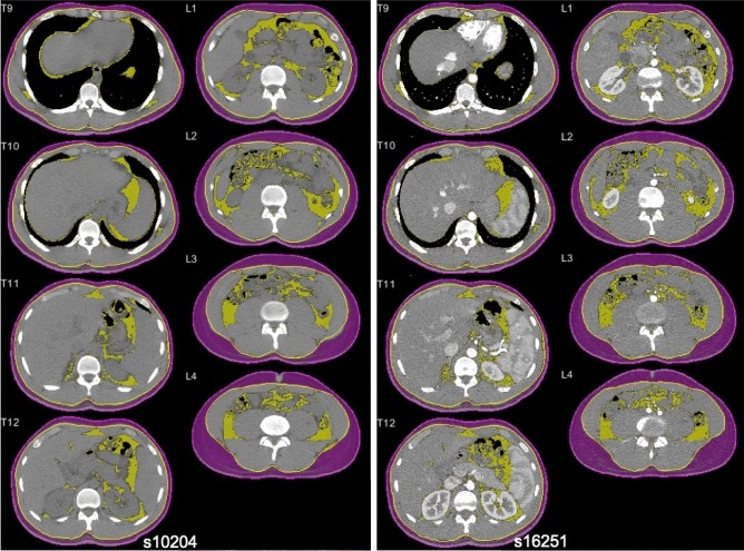
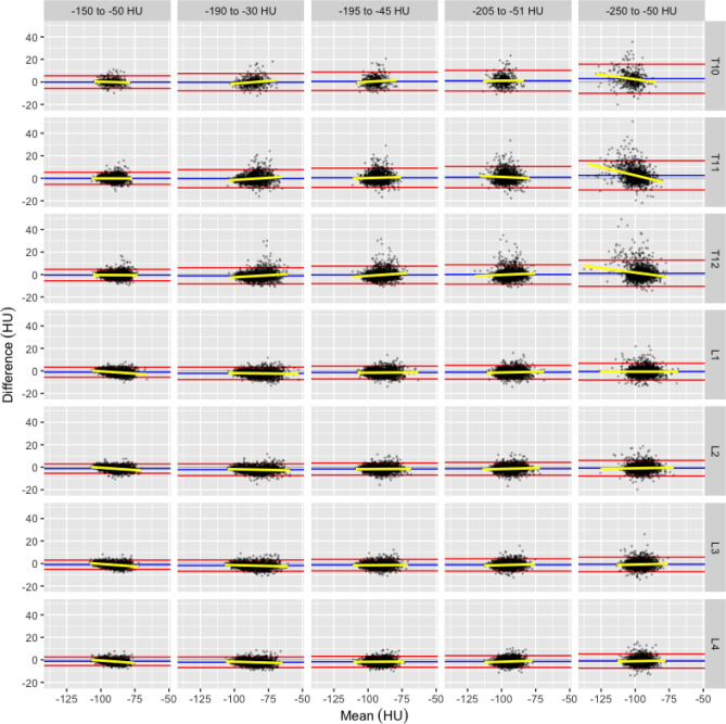
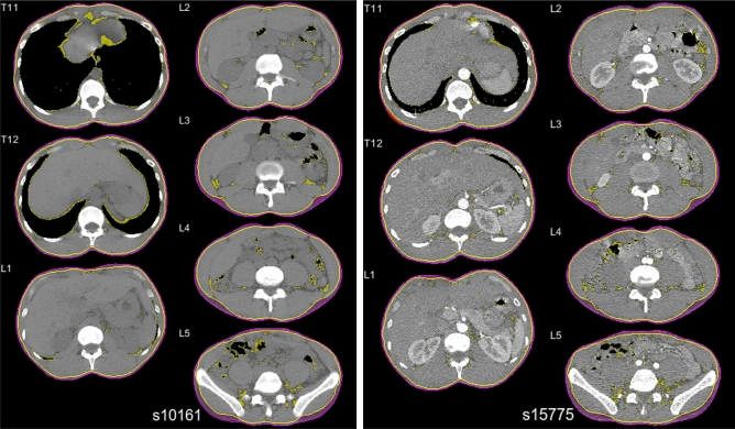
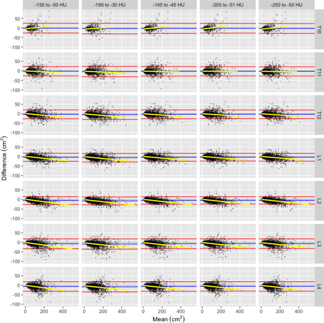
Measurements of visceral adipose tissue cross-sectional area and radiation attenuation from computed tomography (CT) scans provide useful information about risk and mortality. However, scan protocols vary, encompassing differing vertebra levels and utilizing differing phases of contrast enhancement. Furthermore, fat measurements have been extracted from CT using different Hounsfield Unit (HU) ranges. To our knowledge, there have been no large studies of healthy cohorts that reported reference values for visceral fat area and radiation attenuation at multiple vertebra levels, for different contrast phases, and using different fat HU ranges. Two-phase CT scans from 1,677 healthy, adult kidney donors (age 18-65) between 1999 and 2017, previously studied to determine healthy reference values for skeletal muscle measures, were utilized. Visceral adipose tissue cross-sectional area (VFA) and radiation attenuation (VFRA) measures were quantified using axial slices at T10 through L4 vertebra levels. T-tests were used to compare males and females, while paired t-tests were conducted to determine the effect (magnitude and direction) of (a) contrast enhancement and (b) different fat HU ranges on each fat measure at each vertebra level. We report the means, standard deviations, and effect sizes of contrast enhancement and fat HU range. Male and female VFA and VFRA were significantly different at all vertebra levels in both contrast and non-contrast scans. Peak VFA was observed at L4 in females and L2 in males, while peak VFRA was observed at L1 in both females and males. In general, non-contrast scans showed significantly greater VFA and VFRA compared to contrast scans. The average paired difference due to contrast ranged from 1.6 to - 8% (VFA) and 3.2 to - 3.0% (VFRA) of the non-contrast value. HU range showed much greater differences in VFA and VFRA than contrast. The average paired differences due to HU range ranged from - 5.3 to 22.2% (VFA) and - 5.9 to 13.6% (VFRA) in non-contrast scans, and - 4.4 to 20.2% (VFA) and - 4.1 to 12.6% (VFRA) in contrast scans. The - 190 to - 30 HU range showed the largest differences in both VFA (10.8% to 22.2%) and VFRA (7.6% to 13.6%) compared to the reference range (- 205 to - 51 HU). Incidentally, we found that differences in lung inflation result in very large differences in visceral fat measures, particularly in the thoracic region. We assessed the independent effects of contrast presence and fat HU ranges on visceral fat cross-sectional area and mean radiation attenuation, finding significant differences particularly between different fat HU ranges. These results demonstrate that CT measurements of visceral fat area and radiation attenuation are strongly dependent upon contrast presence, fat HU range, sex, breath cycle, and vertebra level of measurement. We quantified contrast and non-contrast reference values separately for males and females, using different fat HU ranges, for lumbar and thoracic CT visceral fat measures at multiple vertebra levels in a healthy adult US population.

https://cdn.ncbi.nlm.nih.gov/pmc/blobs/746e/8837604/05440d36a9ef/41598_2022_6232_Fig4_HTML.jpg
https://cdn.ncbi.nlm.nih.gov/pmc/blobs/746e/8837604/ef4891c46749/41598_2022_6232_Fig1_HTML.jpg
https://cdn.ncbi.nlm.nih.gov/pmc/blobs/746e/8837604/ce04fb35232c/41598_2022_6232_Fig2_HTML.jpg
https://cdn.ncbi.nlm.nih.gov/pmc/blobs/746e/8837604/9965011965b1/41598_2022_6232_Fig3_HTML.jpg
https://cdn.ncbi.nlm.nih.gov/pmc/blobs/746e/8837604/05440d36a9ef/41598_2022_6232_Fig4_HTML.jpg
摘要

从计算机断层扫描(CT)扫描中测量内脏脂肪组织的横截面积和辐射衰减可以提供有关风险和死亡率的有用信息。然而,扫描方案各不相同,包括不同的椎体水平和使用不同的对比增强相位。此外,脂肪测量值是从 CT 中使用不同的 Hounsfield 单位(HU)范围提取的。据我们所知,以前还没有针对健康队列的大型研究报告过在多个椎体水平、不同的对比相和使用不同的脂肪 HU 范围下的内脏脂肪面积和辐射衰减的参考值。我们利用了 1999 年至 2017 年间 1677 名健康成年肾脏供体(年龄 18-65 岁)的两期 CT 扫描,这些扫描先前用于确定骨骼肌测量的健康参考值。使用 T10 至 L4 椎体水平的轴向切片来量化内脏脂肪组织的横截面积(VFA)和辐射衰减(VFRA)。使用 t 检验比较男性和女性,同时进行配对 t 检验以确定(a)对比增强和(b)不同的脂肪 HU 范围对每个脂肪测量值在每个椎体水平上的影响(幅度和方向)。我们报告了对比增强和脂肪 HU 范围的平均值、标准差和效应大小。在男性和女性的两期 CT 扫描中,所有椎体水平的 VFA 和 VFRA 均存在显著差异。女性的 VFA 峰值出现在 L4 椎体,男性的 VFA 峰值出现在 L2 椎体,而女性和男性的 VFRA 峰值均出现在 L1 椎体。通常,非对比扫描显示的 VFA 和 VFRA 明显大于对比扫描。由于对比引起的平均配对差异范围为非对比值的 1.6%至-8%(VFA)和 3.2%至-3.0%(VFRA)。HU 范围在 VFA 和 VFRA 方面显示出比对比更大的差异。由于 HU 范围引起的平均配对差异范围为非对比扫描中的-5.3%至 22.2%(VFA)和-5.9%至 13.6%(VFRA),以及对比扫描中的-4.4%至 20.2%(VFA)和-4.1%至 12.6%(VFRA)。与参考范围(-205 至-51 HU)相比,-190 至-30 HU 范围在 VFA(10.8%至 22.2%)和 VFRA(7.6%至 13.6%)方面显示出最大的差异。顺便说一句,我们发现肺充气的差异会导致内脏脂肪测量值出现非常大的差异,特别是在胸部区域。我们评估了对比剂存在和脂肪 HU 范围对内脏脂肪横截面积和平均辐射衰减的独立影响,发现了特别是在不同脂肪 HU 范围之间的显著差异。这些结果表明,CT 测量的内脏脂肪面积和辐射衰减强烈依赖于对比剂的存在、脂肪 HU 范围、性别、呼吸周期和测量的椎体水平。我们分别使用不同的脂肪 HU 范围,针对男性和女性,在健康的美国成年人中,在多个椎体水平对腰椎和胸部 CT 内脏脂肪测量值进行了非对比和对比参考值的量化。

相似文献

1
Healthy US population reference values for CT visceral fat measurements and the impact of IV contrast, HU range, and spinal levels.美国健康人群 CT 内脏脂肪测量的参考值,以及静脉对比剂、HU 范围和脊柱水平的影响。
Sci Rep. 2022 Feb 11;12(1):2374. doi: 10.1038/s41598-022-06232-5.
2
Relative muscle indices and healthy reference values for sarcopenia assessment using T10 through L5 computed tomography skeletal muscle area.使用 T10 至 L5 计算机断层扫描骨骼肌面积评估肌肉减少症的相对肌肉指数和健康参考值。
Sci Rep. 2024 Sep 18;14(1):21799. doi: 10.1038/s41598-024-71613-x.
3
Visceral adiposity and inflammatory bowel disease.内脏肥胖与炎症性肠病。
Int J Colorectal Dis. 2021 Nov;36(11):2305-2319. doi: 10.1007/s00384-021-03968-w. Epub 2021 Jun 9.
4
Skeletal muscle cutoff values for sarcopenia diagnosis using T10 to L5 measurements in a healthy US population.使用 T10 至 L5 测量值诊断健康美国人群肌少症的骨骼肌截断值。
Sci Rep. 2018 Jul 27;8(1):11369. doi: 10.1038/s41598-018-29825-5.
5
Quantifying Sarcopenia Reference Values Using Lumbar and Thoracic Muscle Areas in a Healthy Population.使用健康人群腰椎和胸椎肌肉面积量化肌少症参考值。
J Nutr Health Aging. 2017;21(10):180-185. doi: 10.1007/s12603-017-0983-3.
6
Correlation between Non-Alcoholic Fatty Liver Disease and Visceral Adipose Tissue in Non-Obese Chinese Adults: A CT Evaluation.非肥胖中国成年人非酒精性脂肪肝与内脏脂肪组织的相关性:CT 评估。
Korean J Radiol. 2018 Sep-Oct;19(5):923-929. doi: 10.3348/kjr.2018.19.5.923. Epub 2018 Aug 6.
7
Effect of contrast phase on quantitative analysis of skeletal muscle and adipose tissue by computed tomography.CT 检查对比相位对骨骼肌和脂肪组织定量分析的影响。
Nutrition. 2024 Oct;126:112492. doi: 10.1016/j.nut.2024.112492. Epub 2024 May 6.
8
Age-related changes in muscle quality and development of diagnostic cutoff points for myosteatosis in lumbar skeletal muscles measured by CT scan.CT 扫描测量腰椎骨骼肌的肌肉质量和肌内脂肪含量诊断切点的年龄相关性变化。
Clin Nutr. 2021 Jun;40(6):4022-4028. doi: 10.1016/j.clnu.2021.04.017. Epub 2021 Apr 17.
9
Diagnostic efficacy of routine contrast-enhanced abdominal CT for the assessment of osteoporosis in the Turkish population.常规增强腹部 CT 诊断效能评估土耳其人群骨质疏松症。
Turk J Med Sci. 2020 Feb 13;50(1):110-116. doi: 10.3906/sag-1906-34.
10
Opportunistic Osteoporosis Screening at Routine Abdominal and Thoracic CT: Normative L1 Trabecular Attenuation Values in More than 20 000 Adults.在常规腹部和胸部 CT 检查中进行机会性骨质疏松症筛查:20000 多名成年人的 L1 小梁衰减正常值。
Radiology. 2019 May;291(2):360-367. doi: 10.1148/radiol.2019181648. Epub 2019 Mar 26.

引用本文的文献

1
The impact of intravenous iodinated contrast agents on radiotherapy dose calculation and radiobiological effect predictions in central lung cancer.静脉注射碘化造影剂对中央型肺癌放疗剂量计算及放射生物学效应预测的影响。
Front Oncol. 2025 Aug 11;15:1563919. doi: 10.3389/fonc.2025.1563919. eCollection 2025.
2
Opportunistic Screening on Chest CT, From the Special Series on Screening.胸部CT的机会性筛查,来自筛查专题系列。
AJR Am J Roentgenol. 2025 Jul 16. doi: 10.2214/AJR.25.33069.
3
Role of tumor-specific and whole-body imaging biomarkers for prediction of recurrence in patients with stage III colorectal cancer.

本文引用的文献

1
3D-segmentation and characterization of visceral and abdominal subcutaneous adipose tissue on CT: influence of contrast medium and contrast phase.CT上内脏和腹部皮下脂肪组织的三维分割与特征分析:造影剂和造影期相的影响
Quant Imaging Med Surg. 2021 Feb;11(2):697-705. doi: 10.21037/qims-20-907.
2
Fully automated CT imaging biomarkers of bone, muscle, and fat: correcting for the effect of intravenous contrast.全自动 CT 成像骨、肌肉和脂肪标志物:校正静脉内对比剂的影响。
Abdom Radiol (NY). 2021 Mar;46(3):1229-1235. doi: 10.1007/s00261-020-02755-5. Epub 2020 Sep 18.
3
Utilizing Fully Automated Abdominal CT-Based Biomarkers for Opportunistic Screening for Metabolic Syndrome in Adults Without Symptoms.
肿瘤特异性和全身成像生物标志物在预测III期结直肠癌患者复发中的作用。
Abdom Radiol (NY). 2025 May;50(5):1907-1915. doi: 10.1007/s00261-024-04656-3. Epub 2024 Nov 2.
4
Incorporating adipose tissue into a CT-based deep learning nomogram to differentiate granulomas from lung adenocarcinomas.将脂肪组织纳入基于CT的深度学习列线图以鉴别肉芽肿与肺腺癌。
iScience. 2024 Aug 19;27(10):110733. doi: 10.1016/j.isci.2024.110733. eCollection 2024 Oct 18.
5
Visceral fat area and subcutaneous fat area as measures of body composition in soft tissue sarcoma.内脏脂肪面积和皮下脂肪面积作为软组织肉瘤身体成分的测量指标。
J Surg Oncol. 2024 Sep;130(3):543-551. doi: 10.1002/jso.27751. Epub 2024 Jun 21.
6
Effects of Interrupting Prolonged Sitting with Light-Intensity Physical Activity on Inflammatory and Cardiometabolic Risk Markers in Young Adults with Overweight and Obesity: Secondary Outcome Analyses of the SED-ACT Randomized Controlled Crossover Trial.中断久坐并进行低强度体力活动对超重和肥胖的年轻成年人的炎症和心血管代谢风险标志物的影响:SED-ACT 随机对照交叉试验的次要结局分析。
Biomolecules. 2024 Aug 19;14(8):1029. doi: 10.3390/biom14081029.
7
Effect of abdominal adipose tissue glucose uptake on brain aging.腹部脂肪组织葡萄糖摄取对大脑老化的影响。
Alzheimers Dement. 2024 Oct;20(10):7104-7112. doi: 10.1002/alz.14193. Epub 2024 Aug 13.
8
Epicardial adipose tissue volume highly correlates with left ventricular diastolic dysfunction in endogenous Cushing's syndrome.心外膜脂肪组织体积与内源性库欣综合征患者的左心室舒张功能障碍高度相关。
Ann Med. 2024 Dec;56(1):2387302. doi: 10.1080/07853890.2024.2387302. Epub 2024 Aug 5.
9
Subcutaneous and Visceral Adipose Tissue Reference Values From the Framingham Heart Study Thoracic and Abdominal CT.来自弗雷明汉心脏研究胸部和腹部CT的皮下及内脏脂肪组织参考值。
Invest Radiol. 2025 Feb 1;60(2):95-104. doi: 10.1097/RLI.0000000000001104. Epub 2024 Jul 25.
10
CT Attenuation of Periaortic Adipose Tissue in Abdominal Aortic Aneurysms.腹主动脉瘤中主动脉周脂肪组织的 CT 衰减。
Radiol Cardiothorac Imaging. 2024 Feb;6(1):e230250. doi: 10.1148/ryct.230250.
利用基于腹部CT的全自动生物标志物对无症状成年人进行代谢综合征的机会性筛查。
AJR Am J Roentgenol. 2021 Jan;216(1):85-92. doi: 10.2214/AJR.20.23049. Epub 2020 Nov 10.
4
Body composition predicts mortality and decompensation in compensated cirrhosis patients: A prospective cohort study.身体成分可预测代偿期肝硬化患者的死亡率和失代偿情况:一项前瞻性队列研究。
JHEP Rep. 2019 Dec 5;2(1):100061. doi: 10.1016/j.jhepr.2019.11.005. eCollection 2020 Feb.
5
Visceral Adipose Tissue Radiodensity Is Linked to Prognosis in Hepatocellular Carcinoma Patients Treated with Selective Internal Radiation Therapy.内脏脂肪组织的放射密度与接受选择性内放射治疗的肝细胞癌患者的预后相关。
Cancers (Basel). 2020 Feb 4;12(2):356. doi: 10.3390/cancers12020356.
6
Body composition evaluation with computed tomography: Contrast media and slice thickness cause methodological errors.体成分评估的 CT 检查:对比剂和层厚可致方法学错误。
Nutrition. 2019 Mar;59:50-55. doi: 10.1016/j.nut.2018.08.001. Epub 2018 Aug 9.
7
Skeletal muscle cutoff values for sarcopenia diagnosis using T10 to L5 measurements in a healthy US population.使用 T10 至 L5 测量值诊断健康美国人群肌少症的骨骼肌截断值。
Sci Rep. 2018 Jul 27;8(1):11369. doi: 10.1038/s41598-018-29825-5.
8
Influence of Contrast Administration on Computed Tomography-Based Analysis of Visceral Adipose and Skeletal Muscle Tissue in Clear Cell Renal Cell Carcinoma.对比剂给药对透明细胞肾细胞癌基于 CT 的内脏脂肪和骨骼肌组织分析的影响。
JPEN J Parenter Enteral Nutr. 2018 Sep;42(7):1148-1155. doi: 10.1002/jpen.1067. Epub 2018 Jan 19.
9
Quantifying Sarcopenia Reference Values Using Lumbar and Thoracic Muscle Areas in a Healthy Population.使用健康人群腰椎和胸椎肌肉面积量化肌少症参考值。
J Nutr Health Aging. 2017;21(10):180-185. doi: 10.1007/s12603-017-0983-3.
10
Percentiles for skeletal muscle index, area and radiation attenuation based on computed tomography imaging in a healthy Caucasian population.基于 CT 成像的健康白种人群骨骼肌指数、面积和辐射衰减百分位数。
Eur J Clin Nutr. 2018 Feb;72(2):288-296. doi: 10.1038/s41430-017-0034-5. Epub 2017 Dec 15.