Song Yi, Wu Qian, Jiang Huojun, Hu Aihao, Xu Lingqi, Tan Caiping, Zhang Biao, Yu Rongming, Qiu Yizhen, Wang Xin, Yang Wenzhong
Department of Critical Care Medicine, Suzhou Hospital of Integrated Traditional Chinese and Western Medicine, Suzhou, China.
Li Shicai School Inheritance Studio, Suzhou Hospital of Integrated Traditional Chinese and Western Medicine, Suzhou, China.
Front Pharmacol. 2022 Jan 26;12:764247. doi: 10.3389/fphar.2021.764247. eCollection 2021.
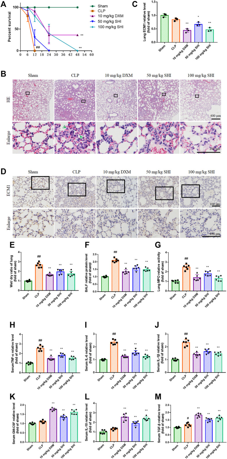
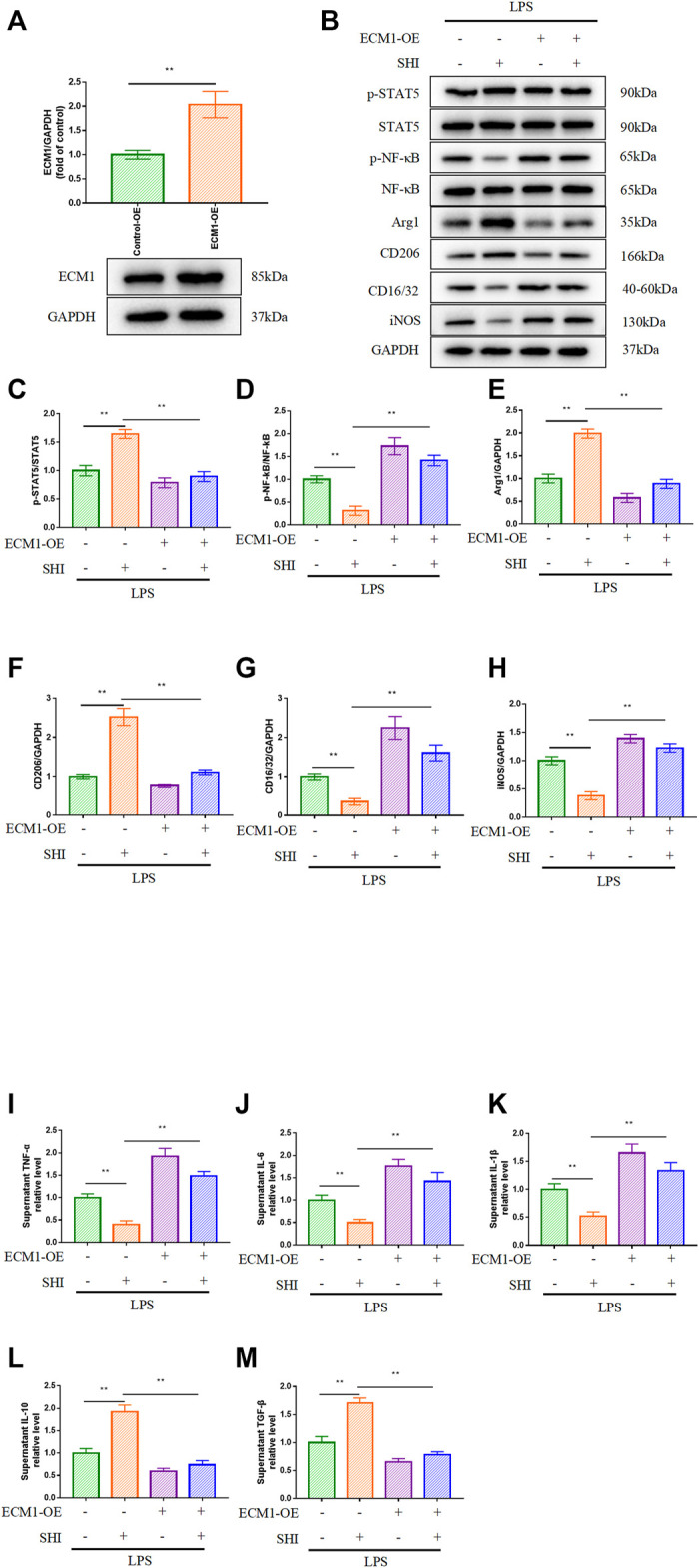
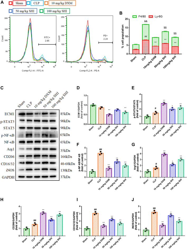
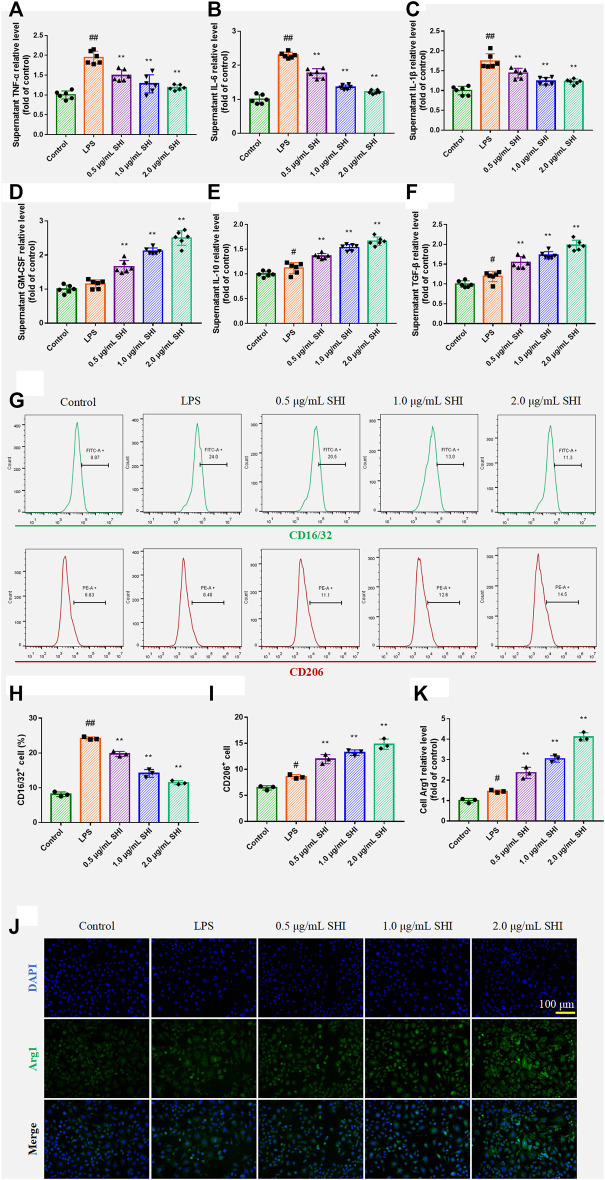
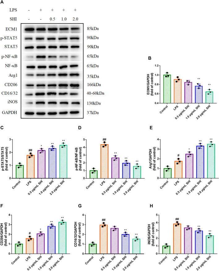
The purpose of the present study was to estimate the effect of shionone (SHI) on sepsis-induced acute lung injury (ALI). The cecal ligation and puncture (CLP) surgery was performed to induce sepsis in mice. Pulmonary hematoxylin and eosin staining, the wet/dry ratio, myeloperoxidase (MPO) activity, and the survival rate were detected. The RAW264.7 cells were treated with SHI and stimulated with lipopolysaccharide (LPS). The cells were also overexpressed by extracellular mechanism protein 1 (ECM1) adenovirus. The relative levels of granulocyte-macrophage colony-stimulating factor, IL-6, IL-1β, TNF-α, IL-10, and TGF-β in the serum and supernatant were measured by ELISA. The protein expressions of ECM1, p-STAT5, signal transducer and activator of transcription 5 (STAT5), p-NF-κB, nuclear factor kappa-B (NF-κB), Arg1, CD206, CD16/32, and iNOS in the CLP-induced lung tissues and LPS-induced cells were detected by western blot. The cell counts of Ly6G, F4/80, CD16/32, and CD206 were evaluated by flow cytometry. The ECM1 expression was also observed by immunohistochemistry and immunofluorescence staining. As a result, the histopathological change, pulmonary edema, and the MPO activity were relieved by SHI. SHI treatment increased the percentage of neutrophil and macrophage in the bronchoalveolar lavage fluid. Besides, SHI administration inhibited pro-inflammatory cytokines and M1 phenotype indices, as well as augmented the anti-inflammatory cytokines and M2 phenotype indices. SHI also attenuated the ECM1/STAT5/NF-κB pathway both and . The overexpression of ECM1 confirmed that the regulated effect of SHI was due to ECM1 signaling. In conclusion, the present study suggests that SHI ameliorated sepsis-induced ALI by screwing M1 phenotype to M2 phenotype macrophage via the ECM1/STAT5/NF-κB pathway.
本研究的目的是评估紫菀酮(SHI)对脓毒症诱导的急性肺损伤(ALI)的影响。通过盲肠结扎和穿刺(CLP)手术诱导小鼠脓毒症。检测肺组织苏木精-伊红染色、湿/干比、髓过氧化物酶(MPO)活性及生存率。用SHI处理RAW264.7细胞并用脂多糖(LPS)刺激。细胞还用细胞外机制蛋白1(ECM1)腺病毒进行过表达。采用酶联免疫吸附测定法(ELISA)检测血清和上清液中粒细胞-巨噬细胞集落刺激因子、白细胞介素-6(IL-6)、白细胞介素-1β(IL-1β)、肿瘤坏死因子-α(TNF-α)、白细胞介素-10(IL-10)和转化生长因子-β(TGF-β)的相对水平。通过蛋白质免疫印迹法检测CLP诱导的肺组织和LPS诱导的细胞中ECM1、磷酸化信号转导和转录激活因子5(p-STAT5)、信号转导和转录激活因子5(STAT5)、磷酸化核因子κB(p-NF-κB)、核因子κB(NF-κB)、精氨酸酶1(Arg1)、CD206、CD16/32和诱导型一氧化氮合酶(iNOS)的蛋白表达。通过流式细胞术评估Ly6G、F4/80、CD16/32和CD206的细胞计数。还通过免疫组织化学和免疫荧光染色观察ECM1表达。结果显示,SHI减轻了组织病理学变化、肺水肿和MPO活性。SHI治疗增加了支气管肺泡灌洗液中中性粒细胞和巨噬细胞的百分比。此外,SHI给药抑制促炎细胞因子和M1表型指标,并增强抗炎细胞因子和M2表型指标。SHI还在体内和体外减弱了ECM1/STAT5/NF-κB通路。ECM1的过表达证实了SHI的调节作用是由于ECM1信号传导。总之,本研究表明,SHI通过ECM1/STAT5/NF-κB通路将M1表型巨噬细胞转变为M2表型巨噬细胞,从而改善脓毒症诱导的ALI。