• 文献检索
  • 文档翻译
  • 深度研究
  • 学术资讯
  • Suppr Zotero 插件Zotero 插件
  • 邀请有礼
  • 套餐&价格
  • 历史记录
应用&插件
Suppr Zotero 插件Zotero 插件浏览器插件Mac 客户端Windows 客户端微信小程序
定价
高级版会员购买积分包购买API积分包
服务
文献检索文档翻译深度研究API 文档MCP 服务
关于我们
关于 Suppr公司介绍联系我们用户协议隐私条款
关注我们

Suppr 超能文献

核心技术专利:CN118964589B侵权必究
粤ICP备2023148730 号-1Suppr @ 2026

文献检索

告别复杂PubMed语法,用中文像聊天一样搜索,搜遍4000万医学文献。AI智能推荐,让科研检索更轻松。

立即免费搜索

文件翻译

保留排版,准确专业,支持PDF/Word/PPT等文件格式,支持 12+语言互译。

免费翻译文档

深度研究

AI帮你快速写综述,25分钟生成高质量综述,智能提取关键信息,辅助科研写作。

立即免费体验

CT肺严重程度评分及定量胸部CT对COVID-19患者分层的诊断性能

Diagnostic performance of CT lung severity score and quantitative chest CT for stratification of COVID-19 patients.

作者信息

Caruso Damiano, Zerunian Marta, Polici Michela, Pucciarelli Francesco, Guido Gisella, Polidori Tiziano, Rucci Carlotta, Bracci Benedetta, Tremamunno Giuseppe, Laghi Andrea

机构信息

Department of Medical Surgical Sciences and Translational Medicine, Sapienza University of Rome, Sant'Andrea University Hospital, Via di Grottarossa, 1035-1039, 00189, Rome, Italy.

出版信息

Radiol Med. 2022 Mar;127(3):309-317. doi: 10.1007/s11547-022-01458-9. Epub 2022 Feb 14.

DOI:10.1007/s11547-022-01458-9
PMID:35157241
原文链接:https://pmc.ncbi.nlm.nih.gov/articles/PMC8852873/
Abstract

PURPOSE

Lung severity score (LSS) and quantitative chest CT (QCCT) analysis could have a relevant impact to stratify patients affected by COVID-19 pneumonia at the hospital admission. The study aims to assess LSS and QCCT performances in severity stratification of COVID-19 patients.

MATERIALS AND METHODS

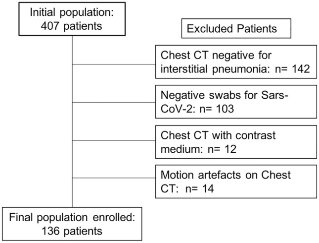
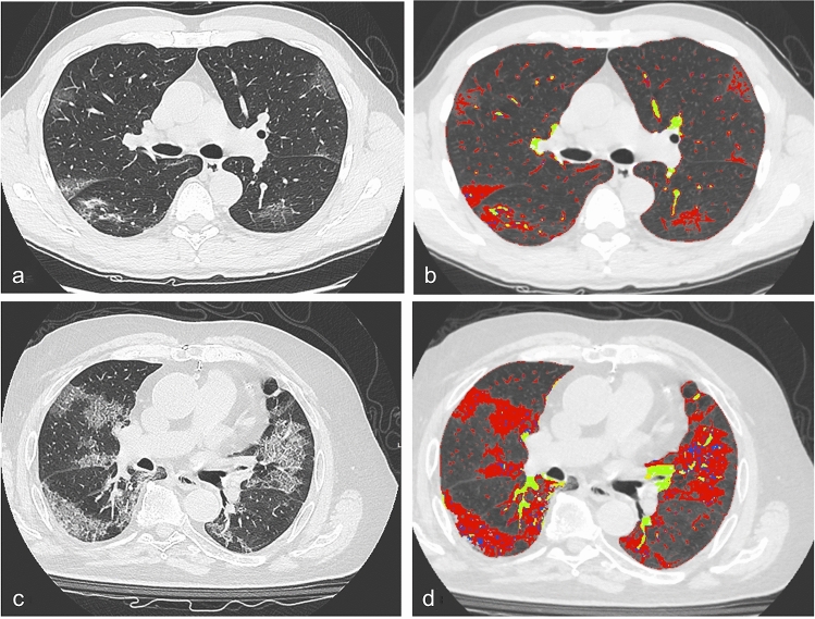
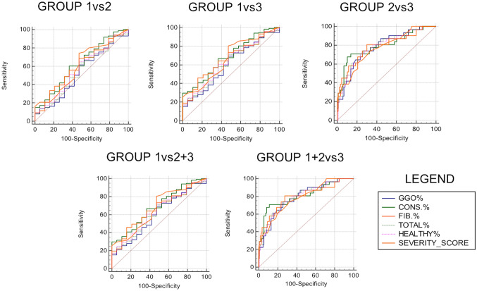
From April 19, 2020, until May 3, 2020, patients with chest CT suggestive for interstitial pneumonia and tested positive for COVID-19 were retrospectively enrolled and stratified for hospital admission as Group 1, 2 and 3 (home isolation, low intensive care and intensive care, respectively). For LSS, lungs were divided in 20 regions and visually assessed by two radiologists who scored for each region from non-lung involvement as 0, < 50% assigned as 1 and > 50% as 2. QCCT was performed with a dedicated software that extracts pulmonary involvement expressed in liters and percentage. LSS and QCCT were analyzed with ROC curve analysis to predict the performance of both methods. P values < 0.05 were considered statistically significant.

RESULTS

Final population enrolled included 136 patients (87 males, mean age 66 ± 16), 19 patients in Group 1, 86 in Group 2 and 31 in Group 3. Significant differences for LSS were observed in almost all comparisons, especially in Group 1 vs 3 (AUC 0.850, P < 0,0001) and Group 1 + 2 vs 3 (AUC 0.783, P < 0,0001). QCCT showed significant results in almost all comparisons, especially between Group 1 vs 3 (AUC 0.869, P < 0,0001). LSS and QCCT comparison between Group 1 and Group 2 did not show significant differences.

CONCLUSIONS

LSS and QCCT could represent promising tools to stratify COVID-19 patient severity at the admission.

摘要

目的

肺部严重程度评分(LSS)和定量胸部CT(QCCT)分析可能对新冠病毒肺炎患者入院时的病情分层产生相关影响。本研究旨在评估LSS和QCCT在新冠病毒患者病情分层中的表现。

材料与方法

从2020年4月19日至2020年5月3日,对胸部CT提示间质性肺炎且新冠病毒检测呈阳性的患者进行回顾性纳入,并在入院时分为1组、2组和3组(分别为居家隔离、低强度监护和重症监护)。对于LSS,将肺部分为20个区域,由两名放射科医生进行视觉评估,他们对每个区域的评分如下:无肺部受累为0分,<50%受累为1分,>50%受累为2分。使用专门软件进行QCCT,该软件可提取以升和百分比表示的肺部受累情况。采用ROC曲线分析对LSS和QCCT进行分析,以预测两种方法的性能。P值<0.05被认为具有统计学意义。

结果

最终纳入的研究对象包括136例患者(87例男性,平均年龄66±16岁),1组19例,2组86例,3组31例。几乎在所有比较中均观察到LSS存在显著差异,尤其是1组与3组(AUC 0.850,P<0.0001)以及1组+2组与3组(AUC 0.783,P<0.0001)。QCCT在几乎所有比较中均显示出显著结果,尤其是1组与3组之间(AUC 0.869,P<0.0001)。1组和2组之间的LSS和QCCT比较未显示出显著差异。

结论

LSS和QCCT可能是入院时对新冠病毒患者病情进行分层的有前景的工具。

https://cdn.ncbi.nlm.nih.gov/pmc/blobs/ac61/8960655/53583cc27ad9/11547_2022_1458_Fig3_HTML.jpg
https://cdn.ncbi.nlm.nih.gov/pmc/blobs/ac61/8960655/c71c6d0ce266/11547_2022_1458_Fig1_HTML.jpg
https://cdn.ncbi.nlm.nih.gov/pmc/blobs/ac61/8960655/42bef3a67bf9/11547_2022_1458_Fig2_HTML.jpg
https://cdn.ncbi.nlm.nih.gov/pmc/blobs/ac61/8960655/53583cc27ad9/11547_2022_1458_Fig3_HTML.jpg
https://cdn.ncbi.nlm.nih.gov/pmc/blobs/ac61/8960655/c71c6d0ce266/11547_2022_1458_Fig1_HTML.jpg
https://cdn.ncbi.nlm.nih.gov/pmc/blobs/ac61/8960655/42bef3a67bf9/11547_2022_1458_Fig2_HTML.jpg
https://cdn.ncbi.nlm.nih.gov/pmc/blobs/ac61/8960655/53583cc27ad9/11547_2022_1458_Fig3_HTML.jpg

相似文献

1
Diagnostic performance of CT lung severity score and quantitative chest CT for stratification of COVID-19 patients.CT肺严重程度评分及定量胸部CT对COVID-19患者分层的诊断性能
Radiol Med. 2022 Mar;127(3):309-317. doi: 10.1007/s11547-022-01458-9. Epub 2022 Feb 14.
2
Post-Acute Sequelae of COVID-19 Pneumonia: Six-month Chest CT Follow-up.新型冠状病毒肺炎的急性后遗症:胸部CT六个月随访
Radiology. 2021 Nov;301(2):E396-E405. doi: 10.1148/radiol.2021210834. Epub 2021 Jul 27.
3
Quantitative Chest CT analysis in discriminating COVID-19 from non-COVID-19 patients.定量胸部 CT 分析在鉴别 COVID-19 与非 COVID-19 患者中的应用。
Radiol Med. 2021 Feb;126(2):243-249. doi: 10.1007/s11547-020-01291-y. Epub 2020 Oct 12.
4
Comparative study of lung ultrasound and chest computed tomography scan in the assessment of severity of confirmed COVID-19 pneumonia.肺超声与胸部 CT 扫描在评估确诊 COVID-19 肺炎严重程度中的对比研究。
Intensive Care Med. 2020 Sep;46(9):1707-1713. doi: 10.1007/s00134-020-06186-0. Epub 2020 Jul 29.
5
Chest CT in the Emergency Department for Diagnosis of COVID-19 Pneumonia: Dutch Experience.急诊科胸部 CT 诊断 COVID-19 肺炎:荷兰经验。
Radiology. 2021 Feb;298(2):E98-E106. doi: 10.1148/radiol.2020203465. Epub 2020 Nov 17.
6
AI-Based Quantitative CT Analysis of Temporal Changes According to Disease Severity in COVID-19 Pneumonia.基于人工智能的 COVID-19 肺炎疾病严重程度与时间变化的定量 CT 分析。
J Comput Assist Tomogr. 2021;45(6):970-978. doi: 10.1097/RCT.0000000000001224.
7
The Performance of Chest CT in Evaluating the Clinical Severity of COVID-19 Pneumonia: Identifying Critical Cases Based on CT Characteristics.胸部 CT 对评估 COVID-19 肺炎临床严重程度的性能:基于 CT 特征识别重症病例。
Invest Radiol. 2020 Jul;55(7):412-421. doi: 10.1097/RLI.0000000000000689.
8
Prognostic Significance of Chest Imaging by LUS and CT in COVID-19 Inpatients: The ECOVID Multicenter Study.超声与 CT 肺部影像学对 COVID-19 住院患者预后评估的意义:ECCOVID 多中心研究。
Respiration. 2022;101(2):122-131. doi: 10.1159/000518516. Epub 2021 Aug 31.
9
Low Sensitivity of Admission Lung US Compared to Chest CT for Diagnosis of Lung Involvement in a Cohort of 82 Patients with COVID-19 Pneumonia.在82例新冠肺炎患者队列中,与胸部CT相比,入院时肺部超声对诊断肺部受累的敏感性较低。
Medicina (Kaunas). 2021 Mar 4;57(3):236. doi: 10.3390/medicina57030236.
10
Computed Tomography Radiomics Can Predict Disease Severity and Outcome in Coronavirus Disease 2019 Pneumonia.计算机断层扫描影像组学可预测2019冠状病毒病肺炎的疾病严重程度和预后。
J Comput Assist Tomogr. 2020 Sep/Oct;44(5):640-646. doi: 10.1097/RCT.0000000000001094.

引用本文的文献

1
Quantitative Chest CT Analysis: Three Different Approaches to Quantify the Burden of Viral Interstitial Pneumonia Using COVID-19 as a Paradigm.胸部CT定量分析:以COVID-19为范例的三种不同方法量化病毒性间质性肺炎的负担
J Clin Med. 2024 Dec 1;13(23):7308. doi: 10.3390/jcm13237308.
2
Lung Involvement in Pulmonary Vasculitis: A Radiological Review.肺部血管炎的肺部受累:影像学综述
Diagnostics (Basel). 2024 Jul 2;14(13):1416. doi: 10.3390/diagnostics14131416.
3
Comparing Visual and Software-Based Quantitative Assessment Scores of Lungs' Parenchymal Involvement Quantification in COVID-19 Patients.

本文引用的文献

1
Recent advances in research on vine tea, a potential and functional herbal tea with dihydromyricetin and myricetin as major bioactive compounds.藤茶是一种具有潜力的功能性草本茶,以二氢杨梅素和杨梅素作为主要生物活性成分,其研究取得了最新进展。
J Pharm Anal. 2021 Oct;11(5):555-563. doi: 10.1016/j.jpha.2020.10.002. Epub 2020 Oct 14.
2
Post-Acute Sequelae of COVID-19 Pneumonia: Six-month Chest CT Follow-up.新型冠状病毒肺炎的急性后遗症:胸部CT六个月随访
Radiology. 2021 Nov;301(2):E396-E405. doi: 10.1148/radiol.2021210834. Epub 2021 Jul 27.
3
Artificial Intelligence for Classification of Soft-Tissue Masses at US.
比较COVID-19患者肺部实质受累量化的视觉和基于软件的定量评估分数
Diagnostics (Basel). 2024 May 8;14(10):985. doi: 10.3390/diagnostics14100985.
4
Serial lung ultrasound in monitoring viral pneumonia: the lesson learned from COVID-19.肺部超声序列监测病毒性肺炎:从新冠疫情中吸取的经验教训
ERJ Open Res. 2023 Aug 14;9(4). doi: 10.1183/23120541.00017-2023. eCollection 2023 Jul.
5
Tips and Tricks in Thoracic Radiology for Beginners: A Findings-Based Approach.初学者胸部放射学技巧与窍门:基于发现的方法。
Tomography. 2023 Jun 14;9(3):1153-1186. doi: 10.3390/tomography9030095.
6
Qualitative and semi-quantitative ultrasound assessment in delta and Omicron Covid-19 patients: data from high volume reference center.德尔塔和奥密克戎新冠患者的定性和半定量超声评估:来自大容量参考中心的数据。
Infect Agent Cancer. 2023 May 27;18(1):34. doi: 10.1186/s13027-023-00515-w.
7
Gender Medicine in Clinical Radiology Practice.临床放射学实践中的性别医学
J Pers Med. 2023 Jan 27;13(2):223. doi: 10.3390/jpm13020223.
8
Radiation Recall Pneumonitis: The Open Challenge in Differential Diagnosis of Pneumonia Induced by Oncological Treatments.放射性回忆性肺炎:肿瘤治疗所致肺炎鉴别诊断中的公开挑战
J Clin Med. 2023 Feb 10;12(4):1442. doi: 10.3390/jcm12041442.
9
Exploring Radiologists' Burnout in the COVID-19 Era: A Narrative Review.探讨 COVID-19 时代放射科医生的职业倦怠:叙事性综述。
Int J Environ Res Public Health. 2023 Feb 14;20(4):3350. doi: 10.3390/ijerph20043350.
用于超声检查中软组织肿块分类的人工智能
Radiol Artif Intell. 2020 Dec 2;3(1):e200125. doi: 10.1148/ryai.2020200125. eCollection 2021 Jan.
4
Combining Initial Radiographs and Clinical Variables Improves Deep Learning Prognostication in Patients with COVID-19 from the Emergency Department.结合初始X光片和临床变量可改善急诊科COVID-19患者的深度学习预后评估。
Radiol Artif Intell. 2020 Dec 16;3(2):e200098. doi: 10.1148/ryai.2020200098. eCollection 2021 Mar.
5
MRI Characteristics, Prevalence, and Outcomes of Hypertrophic Cardiomyopathy with Restrictive Phenotype.具有限制性表型的肥厚型心肌病的MRI特征、患病率及预后
Radiol Cardiothorac Imaging. 2020 Aug 13;2(4):e190158. doi: 10.1148/ryct.2020190158. eCollection 2020 Aug.
6
Differences in Clinical and Imaging Presentation of Pediatric Patients with COVID-19 in Comparison with Adults.新冠病毒病(COVID-19)儿科患者与成人患者在临床和影像学表现上的差异
Radiol Cardiothorac Imaging. 2020 Apr 6;2(2):e200117. doi: 10.1148/ryct.2020200117. eCollection 2020 Apr.
7
Serial Quantitative Chest CT Assessment of COVID-19: A Deep Learning Approach.COVID-19的胸部CT序列定量评估:一种深度学习方法。
Radiol Cardiothorac Imaging. 2020 Mar 30;2(2):e200075. doi: 10.1148/ryct.2020200075. eCollection 2020 Apr.
8
Regulatory Agilities in the Time of COVID-19: Overview, Trends, and Opportunities.《COVID-19 时期的监管动态:概述、趋势与机遇》。
Clin Ther. 2021 Jan;43(1):124-139. doi: 10.1016/j.clinthera.2020.11.015. Epub 2020 Nov 30.
9
Pharmacogenetic considerations in the treatment of co-infections with HIV/AIDS, tuberculosis and malaria in Congolese populations of Central Africa.中非刚果人群中 HIV/AIDS、结核和疟疾合并感染的治疗中的药物遗传学考虑因素。
Int J Infect Dis. 2021 Mar;104:207-213. doi: 10.1016/j.ijid.2020.12.009. Epub 2020 Dec 9.
10
Imaging Changes and Clinical Complications After Drug-Eluting Bead Versus Conventional Transarterial Chemoembolization for Unresectable Hepatocellular Carcinoma: Multicenter Study.不可切除肝细胞癌患者经载药微球与传统经动脉化疗栓塞治疗后的影像学改变及临床并发症:多中心研究。
AJR Am J Roentgenol. 2021 Oct;217(4):933-943. doi: 10.2214/AJR.20.24708. Epub 2020 Nov 27.