Szczepanik-Kułak Paulina, Michalak-Stoma Anna, Krasowska Dorota
Department of Dermatology, Venerology and Paediatric Dermatology, Medical University of Lublin, 20-081 Lublin, Poland.
J Clin Med. 2022 Jan 30;11(3):764. doi: 10.3390/jcm11030764.
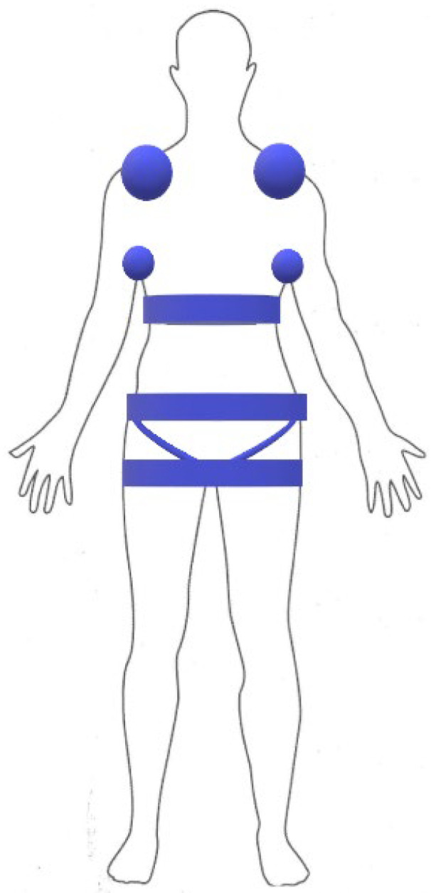
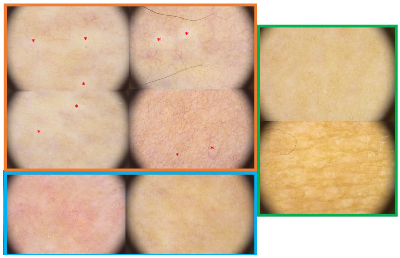
Morphea, also known as localized scleroderma (LoS), is a chronic autoimmune disease of the connective tissue. The clinical picture of LoS distinguishes between active and inactive lesions. Sometimes the clinical findings are challenging to identify, and therefore, the need for additional methods is emphasized. Our study aimed to demonstrate the characteristic dermoscopic features in morphea skin lesions, focusing on demonstrating features in active and inactive lesions. In our patients ( = 31) with histopathologically proven LoS, we performed clinical evaluation of lesions ( = 162): active/inactive and according to both disease activity (modified localized scleroderma severity index, mLoSSI) and damage (localized scleroderma skin damage index, LoSDI) parameters. In addition, we took into account compression locations to determine whether skin trauma, a known etiopathogenetic factor in LoS, affects the dermoscopic pattern of the lesions. We performed a dermoscopy of the lesions, categorizing the images according to the severity within the observed field. We showed that within the active lesions (clinically and with high mLoSSI), white clouds and linear branching vessels had the highest severity. These features decreased within the observed field in inactive lesions and with high LoSDI. Brownish structureless areas were most intense in inactive lesions with high LoSDI. Erythematous areas, linear branching vessels, dotted vessels, and crystalline structures were statistically significant for pressure locations. We have shown dermoscopy is a valuable tool to assess the activity or inactivity of lesions, which translates into appropriate therapeutic decisions and the possibility of monitoring the patient during and after therapy for possible relapse.
硬斑病,也称为局限性硬皮病(LoS),是一种结缔组织的慢性自身免疫性疾病。LoS的临床表现可区分活动期和非活动期病变。有时临床发现难以识别,因此,强调了需要额外的方法。我们的研究旨在展示硬斑病皮肤病变的特征性皮肤镜特征,重点展示活动期和非活动期病变的特征。在我们31例经组织病理学证实为LoS的患者中,我们对162处病变进行了临床评估:根据疾病活动度(改良局限性硬皮病严重程度指数,mLoSSI)和损伤(局限性硬皮病皮肤损伤指数,LoSDI)参数分为活动期/非活动期。此外,我们考虑了压迫部位,以确定皮肤创伤(LoS中已知的病因学因素)是否会影响病变的皮肤镜表现。我们对病变进行了皮肤镜检查,并根据观察视野内的严重程度对图像进行分类。我们发现,在活动期病变(临床和mLoSSI高)中,白云状和线性分支血管的严重程度最高。这些特征在非活动期病变和LoSDI高的病变的观察视野内有所降低。褐色无结构区域在LoSDI高的非活动期病变中最为明显。红斑区域、线性分支血管、点状血管和晶体结构在压迫部位具有统计学意义。我们已经表明,皮肤镜是评估病变活动或非活动的有价值工具,这转化为适当的治疗决策以及在治疗期间和治疗后监测患者是否可能复发的可能性。