Rohowetz Landon J, Mardelli Marc E, Duncan R Scott, Riordan Sean M, Koulen Peter
Department of Ophthalmology, Vision Research Center, School of Medicine, University of Missouri - Kansas City, Kansas City, MO, United States.
Department of Biomedical Sciences, School of Medicine, University of Missouri-Kansas City, Kansas City, MO, United States.
Front Neurosci. 2022 Feb 3;15:801184. doi: 10.3389/fnins.2021.801184. eCollection 2021.
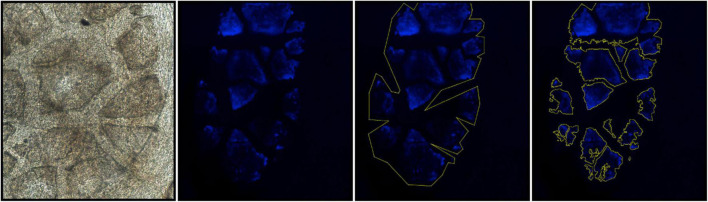
The contributions of anterior segment abnormalities to the development of ocular hypertension was determined in the DBA/2J mouse model of glaucoma. Intraocular pressure (IOP) was measured non-invasively. Iris pigment dispersion (IPD) and corneal calcification were measured weekly starting at 20 weeks of age in DBA/2J and DBA/2J- /SjJ mice. Thickness, surface area, auto-fluorescence intensity, and perimeter length of calcified regions were measured in postmortem corneas using confocal microscopy. DBA/2J mice developed elevated IOP between 9 and 12 months of age, but DBA/2J- /SjJ mice did not. Corneal calcification was found at all ages observed and at similar frequencies in both strains with 83.3% of DBA/2J eyes and 60.0% of DBA/2J- /SjJ eyes affected at 12 months ( = 0.11). Calcification increased with age in both DBA/2J ( = 0.049) and DBA/2J- /SjJ mice ( = 0.04) when assessed qualitatively and based on mixed-effects analysis. No differences in the four objective measures of calcification were observed between strains or ages. At 12 months of age, DBA/2J mice with corneal calcification had greater mean IOP than DBA/2J mice without corneal calcification. IOP was not correlated with the qualitatively assessed measures of calcification. For the subset of eyes with ocular hypertension, which were only found in DBA/2J mice, IOP was negatively correlated with the qualitative degree of calcification, but was not correlated with the four quantitative measures of calcification. Differences in IOP were not observed between DBA/2J- /SjJ mice with and without calcification at any age. IPD increased with age and demonstrated a moderate correlation with IOP in DBA/2J mice, but was not observed in DBA/2J- /SjJ mice. In the DBA/2J mouse model of glaucoma, increased IPD is positively correlated with an increase in IOP and corneal calcification is present in the majority of eyes at and after age 9 months. However, while IPD causes ocular hypertension, corneal calcification does not appear to contribute to the elevation of IOP, as the control strain DBA/2J- /SjJ exhibits corneal calcification similar to DBA/2J mice, but does not develop ocular hypertension. Corneal calcification, therefore, does not appear to be a contributing factor to the development of elevated IOP in DBA/2J mice.
在青光眼的DBA/2J小鼠模型中,确定了眼前节异常对高眼压发展的影响。采用非侵入性方法测量眼压(IOP)。从20周龄开始,每周对DBA/2J和DBA/2J - /SjJ小鼠的虹膜色素播散(IPD)和角膜钙化情况进行测量。使用共聚焦显微镜在死后的角膜中测量钙化区域的厚度、表面积、自发荧光强度和周长。DBA/2J小鼠在9至12月龄时眼压升高,但DBA/2J - /SjJ小鼠未出现这种情况。在观察的所有年龄段均发现了角膜钙化,且两个品系的发生率相似,12月龄时,83.3%的DBA/2J小鼠眼睛和60.0%的DBA/2J - /SjJ小鼠眼睛受到影响(P = 0.11)。定性评估并基于混合效应分析时,DBA/2J小鼠(P = 0.049)和DBA/2J - /SjJ小鼠(P = 0.04)的钙化均随年龄增加。在品系或年龄之间,未观察到钙化的四项客观测量指标存在差异。12月龄时,有角膜钙化的DBA/2J小鼠的平均眼压高于无角膜钙化的DBA/2J小鼠。眼压与定性评估的钙化指标无关。对于仅在DBA/2J小鼠中发现的高眼压眼睛亚组,眼压与定性钙化程度呈负相关,但与钙化的四项定量测量指标无关。在任何年龄,有和无钙化的DBA/2J - /SjJ小鼠之间均未观察到眼压差异。IPD随年龄增加,且在DBA/2J小鼠中与眼压呈中度相关,但在DBA/2J - /SjJ小鼠中未观察到这种情况。在青光眼的DBA/2J小鼠模型中,IPD增加与眼压升高呈正相关,9月龄及之后的大多数眼睛中存在角膜钙化。然而,虽然IPD导致高眼压,但角膜钙化似乎并未导致眼压升高,因为对照品系DBA/2J - /SjJ表现出与DBA/2J小鼠相似的角膜钙化,但未出现高眼压。因此,角膜钙化似乎不是DBA/2J小鼠眼压升高发展的一个促成因素。