Fiedorowicz Michał, Wełniak-Kamińska Marlena, Świątkiewicz Maciej, Orzeł Jarosław, Chorągiewicz Tomasz, Toro Mario Damiano, Rejdak Robert, Bogorodzki Piotr, Grieb Paweł
Department of Experimental Pharmacology, Mossakowski Medical Research Centre, Polish Academy of Sciences, Warsaw, Poland.
Small Animal Magnetic Resonance Imaging Laboratory, Mossakowski Medical Research Centre, Polish Academy of Sciences, Warsaw, Poland.
Front Pharmacol. 2020 Sep 4;11:573238. doi: 10.3389/fphar.2020.573238. eCollection 2020.
The elevation of intraocular pressure (IOP), a major risk factor in glaucoma, is an important parameter tracked in experimental models of this disease. However, IOP measurement in laboratory rodents is challenging and may not correlate with some key pathological events that occur in the development of glaucoma. The aims of this study were to quantify changes in ocular morphology in DBA/2J mice that develop spontaneous, age-dependent, pigmentary glaucoma and to check the possible correlation of these parameters with IOP.
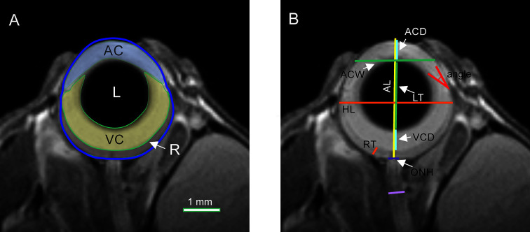
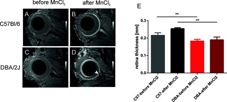
Eye morphology was evaluated with MRI in DBA/2J, DBA/2J-Gpnmb/SjJ, and C57BL/6J female mice ages 3, 6, 9, 12, and 15 months. The animals were anesthetized with isoflurane. A planar receive-only surface coil (inner diameter = 10 mm) was placed over each animal's left eye and the image was acquired with a 7T small animal-dedicated magnetic resonance tomograph and T2-weighted TurboRARE sequence. Ocular dimensions were manually quantitated using OsiriX software. IOP was measured with rebound tonometry.
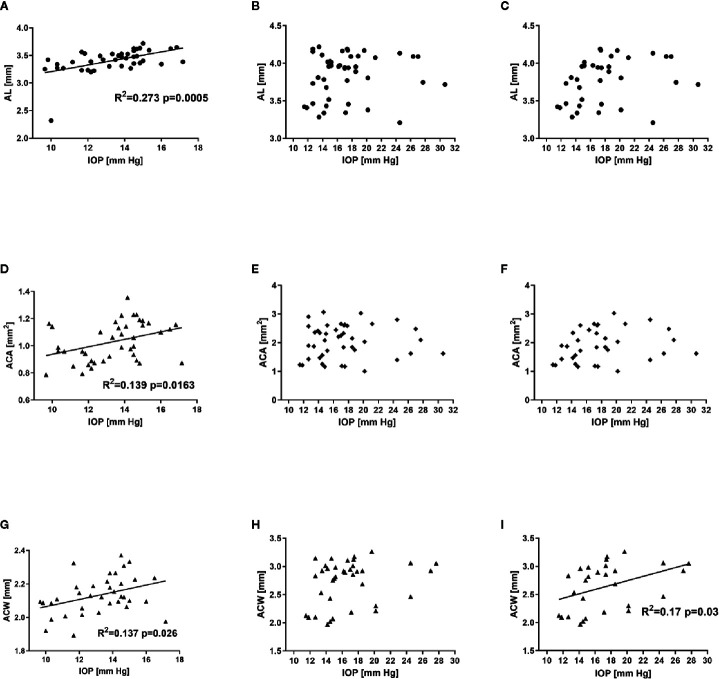
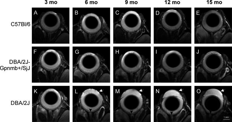
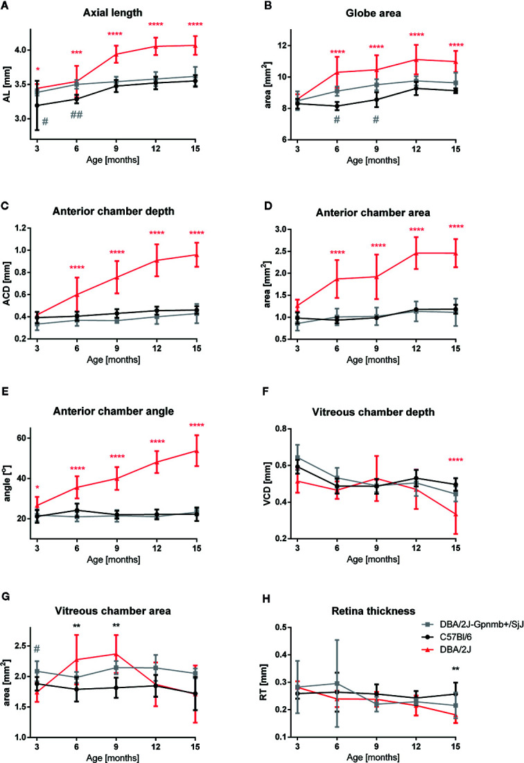
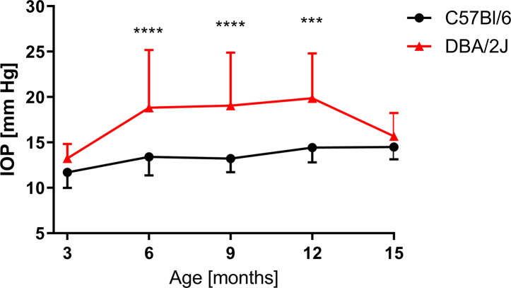
In the control animals, no age-related changes in the ocular morphology were noted. Since 6 months of age, the anterior chamber deepening and elongation of the eyeballs of DBA/2J mice was detectable. We found a significant, positive correlation between IOP and axial length, anterior chamber area, or anterior chamber width in C57BL/6J mice but not in DBA/2J mice. However, after excluding the measurements performed in the oldest DBA/2J mice (i.e. analyzing only the animals ages 3 to 12 months), we demonstrated a significant positive correlation between IOP and anterior chamber width.
High-resolution magnetic resonance imaging of the eye area in mice enables reproducible and consistent measures of key dimensions of the eyeball. We observed age-dependent alterations in the eye morphology of DBA/2J mice that mostly affected the anterior chamber. We also demonstrated a correlation between some of the ocular dimensions and the IOP of C57Bl/6J mice and DBA/2J mice with moderately advanced glaucomatous pathology.
眼压升高是青光眼的主要危险因素,是该疾病实验模型中追踪的重要参数。然而,在实验啮齿动物中测量眼压具有挑战性,且可能与青光眼发展过程中发生的一些关键病理事件不相关。本研究的目的是量化发生自发性、年龄依赖性色素性青光眼的DBA/2J小鼠的眼部形态变化,并检查这些参数与眼压之间的可能相关性。
对3、6、9、12和15月龄的DBA/2J、DBA/2J-Gpnmb/SjJ和C57BL/6J雌性小鼠进行MRI眼部形态评估。动物用异氟醚麻醉。将一个仅用于接收的平面表面线圈(内径 = 10毫米)置于每只动物的左眼上方,并用7T小动物专用磁共振断层扫描仪和T2加权TurboRARE序列采集图像。使用OsiriX软件手动定量眼部尺寸。用回弹眼压计测量眼压。
在对照动物中,未观察到与年龄相关的眼部形态变化。自6月龄起,可检测到DBA/2J小鼠前房加深和眼球伸长。我们发现C57BL/6J小鼠的眼压与眼轴长度、前房面积或前房宽度之间存在显著正相关,但在DBA/2J小鼠中未发现。然而,在排除最年长的DBA/2J小鼠(即仅分析3至12月龄的动物)的测量值后,我们证明眼压与前房宽度之间存在显著正相关。
小鼠眼部区域的高分辨率磁共振成像能够对眼球关键尺寸进行可重复且一致的测量。我们观察到DBA/2J小鼠的眼部形态存在年龄依赖性改变,主要影响前房。我们还证明了C57Bl/6J小鼠和患有中度晚期青光眼病理的DBA/2J小鼠的一些眼部尺寸与眼压之间存在相关性。