Schulz Andreas S, Harteveld Cornelis A M, Vancso G Julius, Huskens Jurriaan, Cloetens Peter, Vos Willem L
Complex Photonic Systems (COPS), MESA+ Institute for Nanotechnology, University of Twente, P.O. Box 217, 7500 AE Enschede, The Netherlands.
Molecular Nanofabrication (MNF), MESA+ Institute for Nanotechnology, University of Twente, P.O. Box 217, 7500 AE Enschede, The Netherlands.
ACS Nano. 2022 Mar 22;16(3):3674-3683. doi: 10.1021/acsnano.1c06915. Epub 2022 Feb 21.
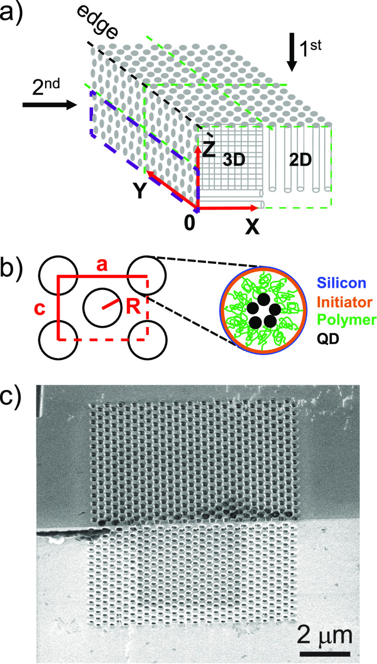
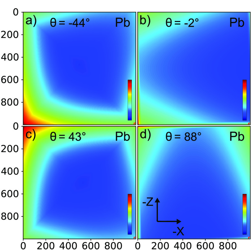
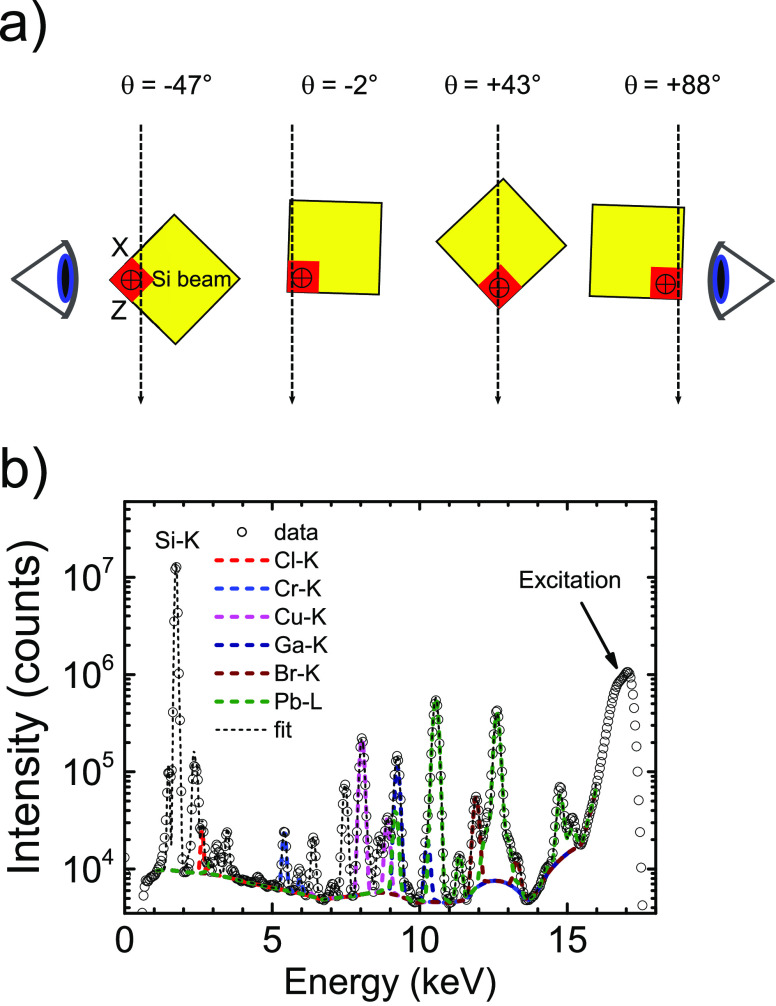
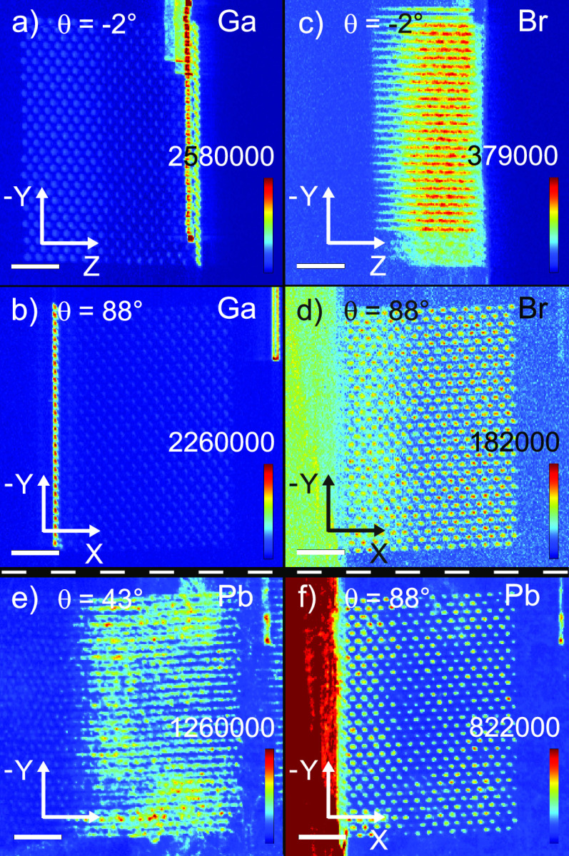
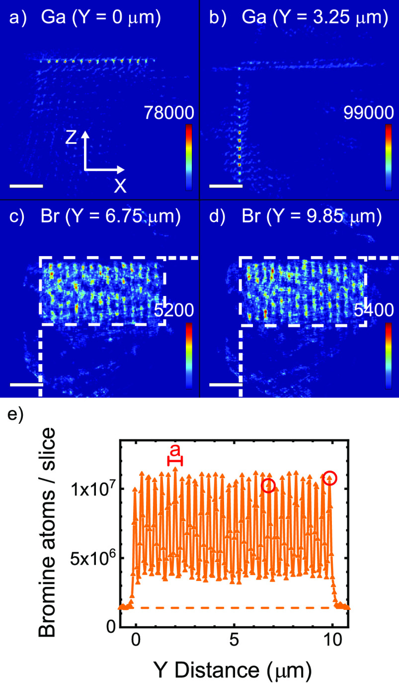
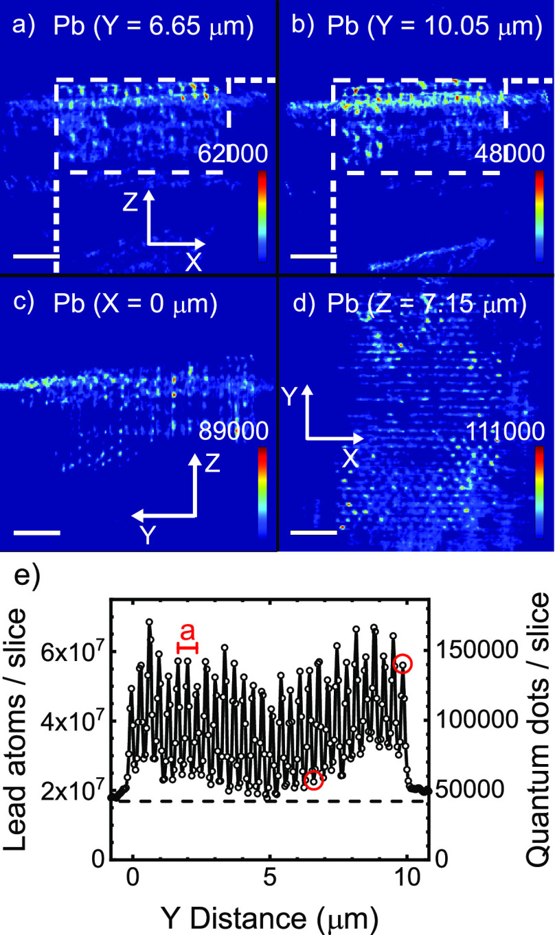
It is a major outstanding goal in nanotechnology to precisely position functional nanoparticles, such as quantum dots, inside a three-dimensional (3D) nanostructure in order to realize innovative functions. Once the 3D positioning is performed, the challenge arises how to nondestructively verify where the nanoparticles reside in the 3D nanostructure. Here, we study 3D photonic band gap crystals made of Si that are infiltrated with PbS nanocrystal quantum dots. The nanocrystals are covalently bonded to polymer brush layers that are grafted to the Si-air interfaces inside the 3D nanostructure using surface-initiated atom transfer radical polymerization (SI-ATRP). The functionalized 3D nanostructures are probed by synchrotron X-ray fluorescence (SXRF) tomography that is performed at 17 keV photon energy to obtain large penetration depths and efficient excitation of the elements of interest. Spatial projection maps were obtained followed by tomographic reconstruction to obtain the 3D atom density distribution with 50 nm voxel size for all chemical elements probed: Cl, Cr, Cu, Ga, Br, and Pb. The quantum dots are found to be positioned inside the 3D nanostructure, and their positions correlate with the positions of elements characteristic of the polymer brush layer and the ATRP initiator. We conclude that X-ray fluorescence tomography is very well suited to nondestructively characterize 3D nanomaterials with photonic and other functionalities.
在纳米技术领域,一个主要的突出目标是将功能纳米粒子(如量子点)精确地定位在三维(3D)纳米结构内部,以实现创新功能。一旦完成3D定位,就会出现如何无损验证纳米粒子在3D纳米结构中所处位置的挑战。在此,我们研究了由硅制成并渗透有硫化铅纳米晶体量子点的3D光子带隙晶体。这些纳米晶体通过表面引发的原子转移自由基聚合(SI-ATRP)与聚合物刷层共价结合,聚合物刷层接枝到3D纳米结构内部的硅-空气界面上。利用在17 keV光子能量下进行的同步加速器X射线荧光(SXRF)断层扫描对功能化的3D纳米结构进行探测,以获得较大的穿透深度并有效激发感兴趣的元素。获得空间投影图,然后进行断层重建,以获得所有探测化学元素(氯、铬、铜、镓、溴和铅)的体素大小为50 nm的3D原子密度分布。发现量子点位于3D纳米结构内部,并且它们的位置与聚合物刷层和ATRP引发剂的特征元素的位置相关。我们得出结论,X射线荧光断层扫描非常适合无损表征具有光子和其他功能的3D纳米材料。