Department of Neurosurgery, Washington University in St. Louis School of Medicine, St. Louis, MO, 63110, USA.
Division of Comparative Medicine, Washington University in St. Louis School of Medicine, St. Louis, MO, 63110, USA.
Fluids Barriers CNS. 2022 Feb 22;19(1):17. doi: 10.1186/s12987-022-00313-3.
Hydrocephalus is a neurological disease with an incidence of 80-125 per 100,000 births in the United States. Neuropathology comprises ventriculomegaly, periventricular white matter (PVWM) alterations, inflammation, and gliosis. We hypothesized that hydrocephalus in a pig model is associated with subventricular and PVWM cellular alterations and neuroinflammation that could mimic the neuropathology described in hydrocephalic infants.
Hydrocephalus was induced by intracisternal kaolin injections in 35-day old female pigs (n = 7 for tissue analysis, n = 10 for CSF analysis). Age-matched sham controls received saline injections (n = 6). After 19-40 days, MRI scanning was performed to measure the ventricular volume. Stem cell proliferation was studied in the Subventricular Zone (SVZ), and cell death and oligodendrocytes were examined in the PVWM. The neuroinflammatory reaction was studied by quantifying astrocytes and microglial cells in the PVWM, and inflammatory cytokines in the CSF.
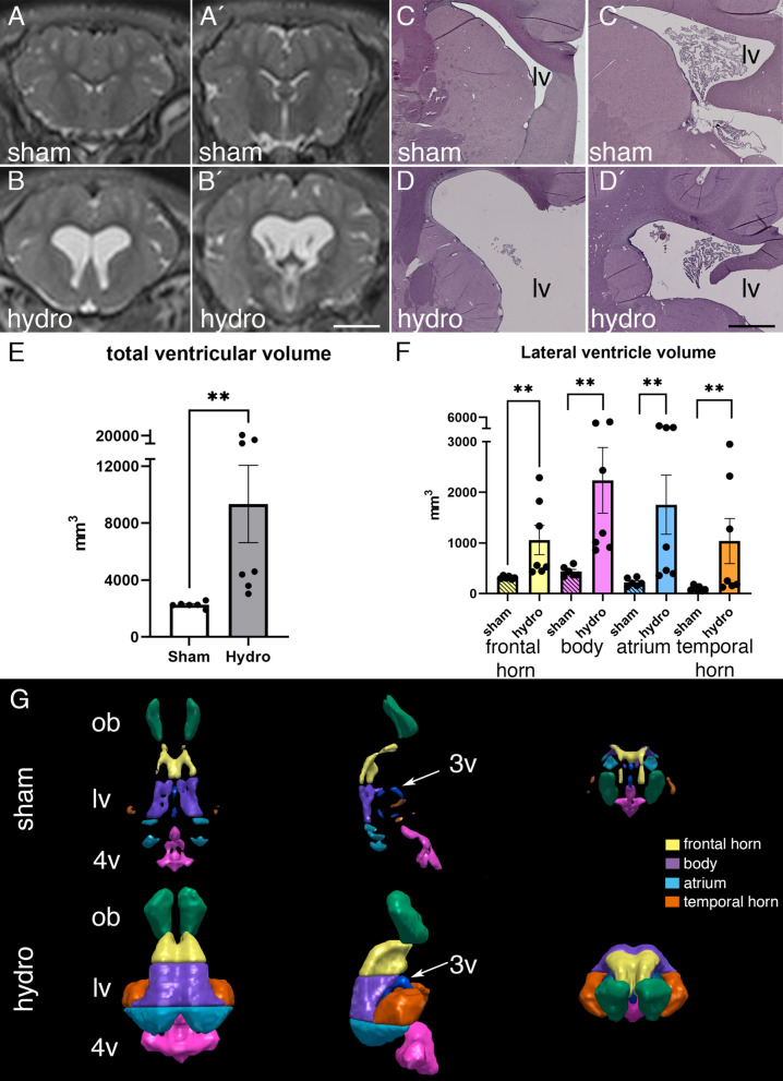
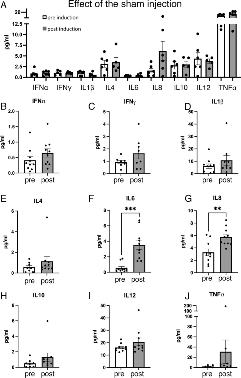
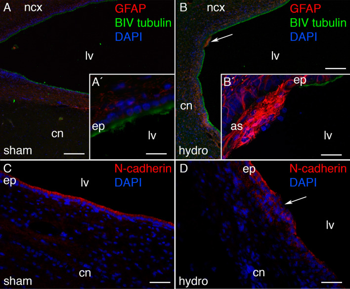
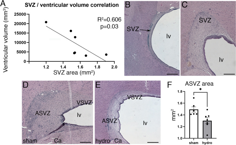
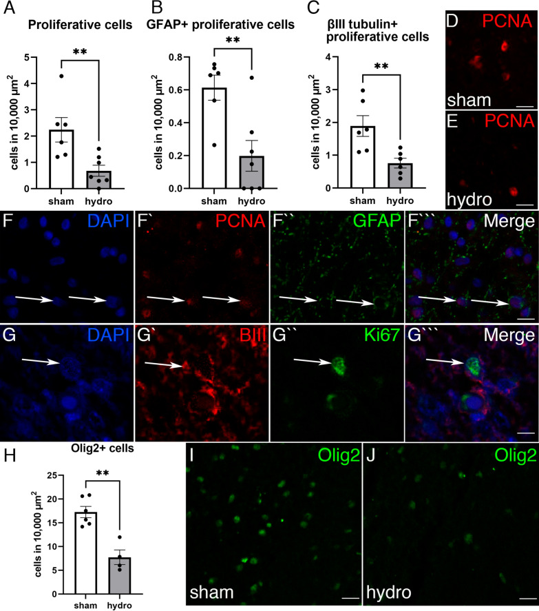
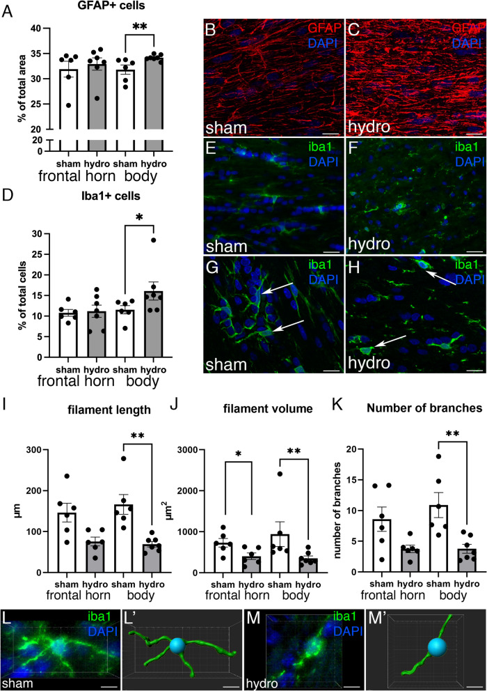
The expansion of the ventricles was especially pronounced in the body of the lateral ventricle, where ependymal disruption occurred. PVWM showed a 44% increase in cell death and a 67% reduction of oligodendrocytes. In the SVZ, the number of proliferative cells and oligodendrocyte decreased by 75% and 57% respectively. The decrease of the SVZ area correlated significantly with ventricular volume increase. Neuroinflammation occurred in the hydrocephalic pigs with a significant increase of astrocytes and microglia in the PVWM, and high levels of inflammatory interleukins IL-6 and IL-8 in the CSF.
The induction of acquired hydrocephalus produced alterations in the PVWM, reduced cell proliferation in the SVZ, and neuroinflammation.
在美国,脑积水的发病率为每 10 万人中有 80-125 例。神经病理学包括脑室扩大、脑室周围白质(PVWM)改变、炎症和神经胶质增生。我们假设猪模型中的脑积水与脑室下区和 PVWM 细胞改变以及神经炎症有关,这些改变可能模拟脑积水婴儿的神经病理学。
通过向 35 天大的雌性猪的脑室内注射高岭土来诱导脑积水(组织分析 n=7,CSF 分析 n=10)。年龄匹配的假手术对照组接受生理盐水注射(n=6)。19-40 天后,进行 MRI 扫描以测量脑室容积。研究了 SVZ 中的干细胞增殖,并在 PVWM 中检查了细胞死亡和少突胶质细胞。通过量化 PVWM 中的星形胶质细胞和小胶质细胞以及 CSF 中的炎症细胞因子来研究神经炎症反应。
脑室的扩张在侧脑室体部尤其明显,那里出现了室管膜破坏。PVWM 中的细胞死亡增加了 44%,少突胶质细胞减少了 67%。在 SVZ 中,增殖细胞和少突胶质细胞的数量分别减少了 75%和 57%。SVZ 面积的减少与脑室容积的增加显著相关。在脑积水猪中发生了神经炎症,PVWM 中的星形胶质细胞和小胶质细胞显著增加,CSF 中的炎症细胞因子 IL-6 和 IL-8 水平升高。
获得性脑积水的诱导导致了 PVWM 的改变、SVZ 中细胞增殖减少和神经炎症。