National Heart and Lung Institute, London, UK.
MRC Asthma UK Centre in Allergic Mechanisms of Asthma, London, UK.
Clin Exp Allergy. 2022 Apr;52(4):550-560. doi: 10.1111/cea.14116. Epub 2022 Mar 3.
Rhinoviruses are the major precipitant of asthma exacerbations and individuals with asthma experience more severe/prolonged rhinovirus infections. Concurrent viral infection and allergen exposure synergistically increase exacerbation risk. Although dendritic cells orchestrate immune responses to both virus and allergen, little is known about their role in viral asthma exacerbations.
To characterize dendritic cell populations present in the lower airways, and to assess whether their numbers are altered in asthma compared to healthy subjects prior to infection and during rhinovirus-16 infection.
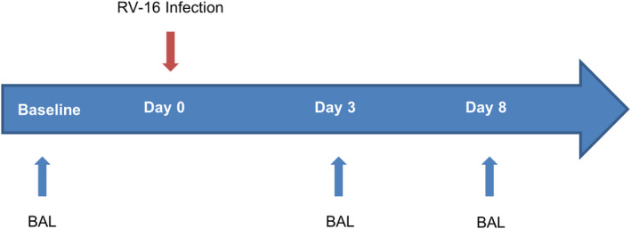
Moderately-severe atopic asthmatic patients and healthy controls were experimentally infected with rhinovirus-16. Bronchoalveolar lavage was collected at baseline, day 3 and day 8 post infection and dendritic cells isolated using fluorescence activated cell sorting.
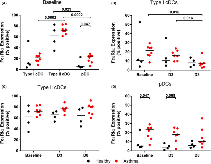
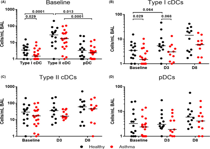
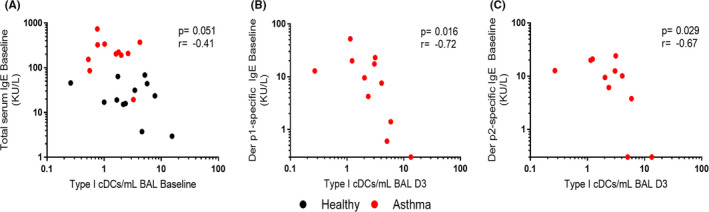
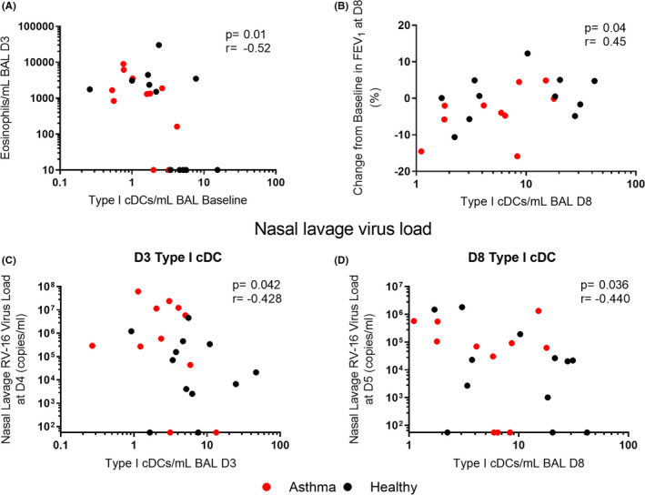
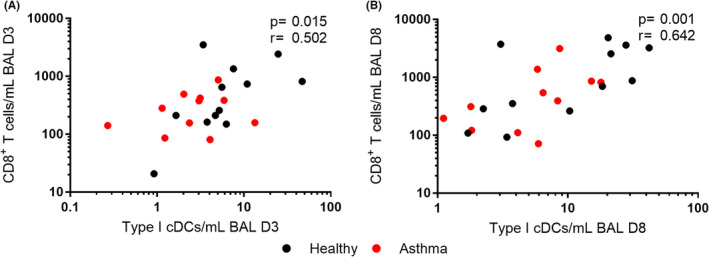
Numbers of type I conventional dendritic cells, which cross prime CD8 T helper cells and produce innate interferons, were significantly reduced in the lower airways of asthma patients compared to healthy controls at baseline. This reduction was associated serum IgE at baseline and with reduced numbers of CD8 T helper cells and with increased viral replication, airway eosinophils and reduced lung function during infection. IgE receptor expression on lower airway plasmacytoid dendritic cells was significantly increased in asthma, consistent with a reduced capacity to produce innate interferons.
Reduced numbers of anti-viral type I conventional dendritic cells in asthma are associated with adverse outcomes during rhinovirus infection. This, with increased FcεR1α expression on lower airway plasmacytoid DCs could mediate the more permissive respiratory viral infection observed in asthma patients.
鼻病毒是哮喘恶化的主要诱因,哮喘患者经历更严重/持久的鼻病毒感染。同时存在病毒感染和过敏原暴露会协同增加恶化风险。尽管树突状细胞协调针对病毒和过敏原的免疫反应,但它们在病毒诱导的哮喘恶化中的作用知之甚少。
描述下呼吸道中存在的树突状细胞群体,并评估与健康受试者相比,在感染前和感染鼻病毒 16 时,哮喘患者的这些细胞数量是否发生改变。
中度严重的特应性哮喘患者和健康对照者经实验性感染鼻病毒 16。在感染前、感染后第 3 天和第 8 天采集支气管肺泡灌洗液,并使用荧光激活细胞分选术分离树突状细胞。
与健康对照组相比,哮喘患者在下呼吸道中的 I 型传统树突状细胞(可交叉呈递 CD8 T 辅助细胞并产生先天干扰素)数量在基线时明显减少。这种减少与基线时的血清 IgE 相关,与 CD8 T 辅助细胞数量减少以及病毒复制增加、气道嗜酸性粒细胞增多和感染期间肺功能降低有关。哮喘患者下呼吸道浆细胞样树突状细胞的 IgE 受体表达显著增加,与先天干扰素产生能力降低一致。
哮喘患者中抗病毒的 I 型传统树突状细胞数量减少与鼻病毒感染期间的不良结局有关。这种情况与下呼吸道浆细胞样树突状细胞上 FcεR1α 表达增加相结合,可能介导了哮喘患者中更易发生的呼吸道病毒感染。