Department of Pathology and Diagnostics and Public Health, Section of Pathology, University of Verona, Verona, Italy.
Department of Pathology and Diagnostics, University of Verona, P.le Stefani n. 1; 37126, Verona, Italy.
Virchows Arch. 2022 Jun;480(6):1223-1230. doi: 10.1007/s00428-022-03304-9. Epub 2022 Feb 25.
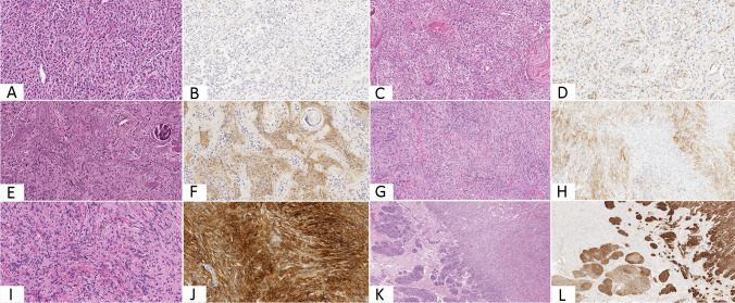
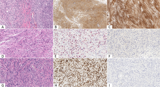
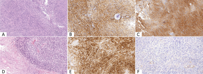
Meningiomas are common tumors of the central nervous system. Although their histological diagnosis is usually straightforward, their differential diagnosis versus other tumors may be challenging at times. The objective of this study is to assess the diagnostic value of CD13 immunoexpression in the differential diagnosis between meningiomas and their morphological mimics. Immunohistochemical analysis for CD13, epithelial membrane antigen, SOX10, and STAT6 was carried out in a large cohort of primary meningeal tumors comprising 225 meningiomas, 15 schwannomas, and 20 solitary fibrous tumor/hemangiopericytomas. Within the meningioma group, the expression of CD13 and epithelial membrane antigen was distinguished in three categories using a semiquantitative score. Most of meningiomas expressed CD13 (94%) and epithelial membrane antigen (96%) while none of the schwannomas nor of the solitary fibrous tumor/hemangiopericytomas was positive for either the two markers. Diffuse positivity for CD13 and epithelial membrane antigen was more common in low-grade meningiomas than in anaplastic ones, which were also more often negative for such markers, especially for CD13 (32%). CD13 is a helpful immunohistochemical marker for the differential diagnosis of meningiomas and their mimics, achieving in combination with epithelial membrane antigen maximal sensitivity (100%) and showing statistically relevant difference of expression in comparison with both schwannomas (p < 0.0001) and solitary fibrous tumor/hemangiopericytomas (p < 0.0001). Furthermore, loss of CD13 expression could be related to outcome as it is associated with worrisome histological findings, mainly in the setting of anaplastic meningiomas.
脑膜瘤是中枢神经系统常见的肿瘤。尽管其组织学诊断通常较为明确,但有时与其他肿瘤的鉴别诊断仍具有挑战性。本研究旨在评估 CD13 免疫表达在脑膜瘤及其形态学模拟物鉴别诊断中的诊断价值。对包括 225 例脑膜瘤、15 例神经鞘瘤和 20 例孤立性纤维瘤/血管外皮细胞瘤在内的原发性脑膜瘤大队列进行了 CD13、上皮膜抗原、SOX10 和 STAT6 的免疫组织化学分析。在脑膜瘤组中,使用半定量评分将 CD13 和上皮膜抗原的表达分为三类。大多数脑膜瘤(94%)和上皮膜抗原(96%)表达 CD13,而神经鞘瘤和孤立性纤维瘤/血管外皮细胞瘤均不表达这两种标志物。低级别脑膜瘤中 CD13 和上皮膜抗原弥漫阳性的比例高于间变性脑膜瘤,后两者也更常为这些标志物阴性,尤其是 CD13(32%)。CD13 是脑膜瘤及其模拟物鉴别诊断的有用免疫组织化学标志物,与上皮膜抗原联合使用可实现最高的敏感性(100%),并且与神经鞘瘤(p<0.0001)和孤立性纤维瘤/血管外皮细胞瘤(p<0.0001)相比,其表达具有统计学上显著差异。此外,CD13 表达缺失可能与预后有关,因为它与令人担忧的组织学发现有关,主要是在间变性脑膜瘤中。