• 文献检索
  • 文档翻译
  • 深度研究
  • 学术资讯
  • Suppr Zotero 插件Zotero 插件
  • 邀请有礼
  • 套餐&价格
  • 历史记录
应用&插件
Suppr Zotero 插件Zotero 插件浏览器插件Mac 客户端Windows 客户端微信小程序
定价
高级版会员购买积分包购买API积分包
服务
文献检索文档翻译深度研究API 文档MCP 服务
关于我们
关于 Suppr公司介绍联系我们用户协议隐私条款
关注我们

Suppr 超能文献

核心技术专利:CN118964589B侵权必究
粤ICP备2023148730 号-1Suppr @ 2026

文献检索

告别复杂PubMed语法,用中文像聊天一样搜索,搜遍4000万医学文献。AI智能推荐,让科研检索更轻松。

立即免费搜索

文件翻译

保留排版,准确专业,支持PDF/Word/PPT等文件格式,支持 12+语言互译。

免费翻译文档

深度研究

AI帮你快速写综述,25分钟生成高质量综述,智能提取关键信息,辅助科研写作。

立即免费体验

基于 CT 的临床Ⅰ期肺腺癌影像学基因组分析:与组织病理学特征和肿瘤学结局的关系。

CT-based Radiogenomic Analysis of Clinical Stage I Lung Adenocarcinoma with Histopathologic Features and Oncologic Outcomes.

机构信息

From the Department of Radiology (R.P., J.A.A., P.G., M.S.G.), Druckenmiller Center for Lung Cancer Research (R.P., K.S.T., N.R., M.S.G., D.R.J.), Thoracic Surgery Service (J.G.C., R.C., J.Z., D.R.J.), Biostatistics Service, Department of Epidemiology and Biostatistics (K.W., K.S.T.), and Department of Pathology (N.R.), Memorial Sloan Kettering Cancer Center, 1275 York Ave, Box 7, New York, NY 10065.

出版信息

Radiology. 2022 Jun;303(3):664-672. doi: 10.1148/radiol.211582. Epub 2022 Mar 1.

DOI:10.1148/radiol.211582
PMID:35230187
原文链接:https://pmc.ncbi.nlm.nih.gov/articles/PMC9131171/
Abstract

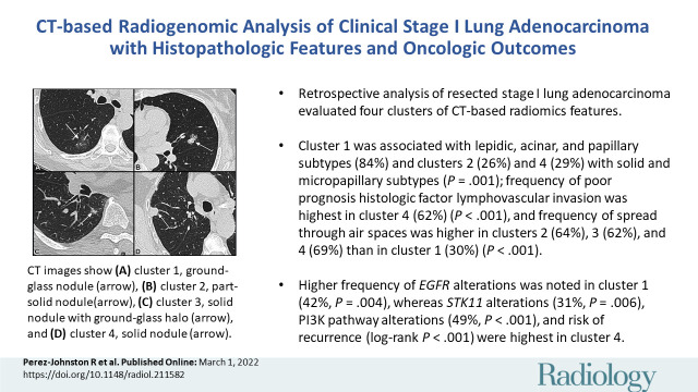
Background A preoperative predictive model is needed that can be used to identify patients with lung adenocarcinoma (LUAD) who have a higher risk of recurrence or metastasis. Purpose To investigate associations between CT-based radiomic consensus clustering of stage I LUAD and clinical-pathologic features, genomic data, and patient outcomes. Materials and Methods Patients who underwent complete surgical resection for LUAD from April 2014 to December 2017 with preoperative CT and next-generation sequencing data were retrospectively identified. Comprehensive radiomic analysis was performed on preoperative CT images; tumors were classified as solid, ground glass, or mixed. Patients were clustered into groups based on their radiomics features using consensus clustering, and clusters were compared with tumor genomic alterations, histopathologic features, and recurrence-specific survival (Kruskal-Wallis test for continuous data, χ or Fisher exact test for categorical data, and log-rank test for recurrence-specific survival). Cluster analysis was performed on the entire cohort and on the solid, ground-glass, and mixed lesion subgroups. Results In total, 219 patients were included in the study (median age, 68 years; interquartile range, 63-74 years; 150 [68%] women). Four radiomic clusters were identified. Cluster 1 was associated with lepidic, acinar, and papillary subtypes (76 of 90 [84%]); clusters 2 (13 of 50 [26%]) and 4 (13 of 45 [29%]) were associated with solid and micropapillary subtypes ( < .001). The alterations were highest in cluster 1 (38 of 90 [42%], = .004). Clusters 2, 3, and 4 were associated with lymphovascular invasion (19 of 50 [38%], 14 of 34 [41%], and 28 of 45 [62%], respectively; < .001) and tumor spread through air spaces (32 of 50 [64%], 21 of 34 [62%], and 31 of 45 [69%], respectively; < .001). alterations (14 of 45 [31%]; = .006), phosphoinositide 3-kinase pathway alterations (22 of 45 [49%], < .001), and risk of recurrence (log-rank < .001) were highest in cluster 4. Conclusion CT-based radiomic consensus clustering enabled identification of associations between radiomic features and clinicalpathologic and genomic features and outcomes in patients with clinical stage I lung adenocarcinoma. © RSNA, 2022 See also the editorial by Nishino in this issue.

摘要

背景 需要一种术前预测模型,用于识别肺腺癌(LUAD)患者,这些患者具有更高的复发或转移风险。目的 探讨 CT 基于放射组学共识聚类的 I 期 LUAD 与临床病理特征、基因组数据和患者结局之间的关联。材料与方法 回顾性分析 2014 年 4 月至 2017 年 12 月期间因 LUAD 行完全手术切除且术前有 CT 和下一代测序数据的患者。对术前 CT 图像进行全面的放射组学分析;肿瘤分为实性、磨玻璃样或混合性。根据放射组学特征对患者进行共识聚类分组,并对聚类与肿瘤基因组改变、组织病理学特征和复发特异性生存进行比较(连续数据的 Kruskal-Wallis 检验、分类数据的 χ 检验或 Fisher 确切检验、以及复发特异性生存的对数秩检验)。对整个队列和实性、磨玻璃样和混合性病变亚组进行聚类分析。结果 共纳入 219 例患者(中位年龄,68 岁;四分位间距,63-74 岁;150 [68%] 例女性)。确定了 4 个放射组学聚类。聚类 1 与贴壁、腺泡和乳头亚型相关(76/90 [84%]);聚类 2(50/50 [26%])和聚类 4(45/45 [29%])与实性和微乳头亚型相关( <.001)。聚类 1 的 改变率最高(38/90 [42%], =.004)。聚类 2、3 和 4 与脉管侵犯(50/50 [64%]、34/34 [41%]和 45/45 [62%], <.001)和肿瘤通过气腔播散(50/50 [64%]、34/34 [62%]和 45/45 [69%], <.001)相关。聚类 4 的 改变率(14/45 [31%]; =.006)、磷酸肌醇 3-激酶通路改变率(22/45 [49%], <.001)和复发风险(对数秩 <.001)最高。结论 CT 基于放射组学的共识聚类可识别临床 I 期肺腺癌患者的放射组学特征与临床病理和基因组特征及结局之间的关联。

https://cdn.ncbi.nlm.nih.gov/pmc/blobs/cda6/9131171/4455abab7f73/radiol.211582.VA.jpg

相似文献

1
CT-based Radiogenomic Analysis of Clinical Stage I Lung Adenocarcinoma with Histopathologic Features and Oncologic Outcomes.基于 CT 的临床Ⅰ期肺腺癌影像学基因组分析:与组织病理学特征和肿瘤学结局的关系。
Radiology. 2022 Jun;303(3):664-672. doi: 10.1148/radiol.211582. Epub 2022 Mar 1.
2
Diagnosis of Invasive Lung Adenocarcinoma Based on Chest CT Radiomic Features of Part-Solid Pulmonary Nodules: A Multicenter Study.基于部分实性肺结节 CT 放射组学特征对浸润性肺腺癌的诊断:一项多中心研究。
Radiology. 2020 Nov;297(2):451-458. doi: 10.1148/radiol.2020192431. Epub 2020 Aug 25.
3
A combination of radiomic features, clinic characteristics, and serum tumor biomarkers to predict the possibility of the micropapillary/solid component of lung adenocarcinoma.联合放射组学特征、临床特征和血清肿瘤标志物预测肺腺癌微乳头/实性成分的可能性。
Ther Adv Respir Dis. 2024 Jan-Dec;18:17534666241249168. doi: 10.1177/17534666241249168.
4
Histopathologic Basis for a Chest CT Deep Learning Survival Prediction Model in Patients with Lung Adenocarcinoma.基于胸部 CT 深度学习预测肺腺癌患者生存模型的组织病理学基础。
Radiology. 2022 Nov;305(2):441-451. doi: 10.1148/radiol.213262. Epub 2022 Jul 5.
5
Feasibility of UTE-MRI-based radiomics model for prediction of histopathologic subtype of lung adenocarcinoma: in comparison with CT-based radiomics model.基于 UTE-MRI 的放射组学模型预测肺腺癌组织病理亚型的可行性:与 CT 基于放射组学模型的比较。
Eur Radiol. 2024 May;34(5):3422-3430. doi: 10.1007/s00330-023-10302-1. Epub 2023 Oct 16.
6
CT-based radiomic phenotypes of lung adenocarcinoma: a preliminary comparative analysis with targeted next-generation sequencing.基于CT的肺腺癌放射组学特征:与靶向二代测序的初步对比分析
Front Med (Lausanne). 2023 Aug 17;10:1191019. doi: 10.3389/fmed.2023.1191019. eCollection 2023.
7
Prediction of solid and micropapillary components in lung invasive adenocarcinoma: radiomics analysis from high-spatial-resolution CT data with 1024 matrix.肺浸润性腺癌实性和微乳头状成分的预测:高空间分辨率 CT 数据的 1024 矩阵的放射组学分析。
Jpn J Radiol. 2024 Jun;42(6):590-598. doi: 10.1007/s11604-024-01534-2. Epub 2024 Feb 28.
8
Consensus Clustering Analysis Based on Enhanced-CT Radiomic Features: Esophageal Squamous Cell Carcinoma patients' 3-Year Progression-Free Survival.基于增强 CT 放射组学特征的共识聚类分析:食管鳞状细胞癌患者 3 年无进展生存率。
Acad Radiol. 2024 Jul;31(7):2807-2817. doi: 10.1016/j.acra.2023.12.025. Epub 2024 Jan 9.
9
A novel radiomic nomogram for predicting epidermal growth factor receptor mutation in peripheral lung adenocarcinoma.一种用于预测周围型肺腺癌表皮生长因子受体突变的新型放射组学列线图。
Phys Med Biol. 2020 Mar 6;65(5):055012. doi: 10.1088/1361-6560/ab6f98.
10
Utility of CT radiomics for prediction of PD-L1 expression in advanced lung adenocarcinomas.CT 放射组学预测晚期肺腺癌 PD-L1 表达的效用。
Thorac Cancer. 2020 Apr;11(4):993-1004. doi: 10.1111/1759-7714.13352. Epub 2020 Feb 11.

引用本文的文献

1
Consensus clustering based on CT radiomics has potential for risk stratification of patients with clinical T1 stage lung adenocarcinoma.基于CT影像组学的一致性聚类在临床T1期肺腺癌患者的风险分层中具有潜力。
BMC Med Imaging. 2025 Jul 1;25(1):231. doi: 10.1186/s12880-025-01795-x.
2
Progress and challenges of artificial intelligence in lung cancer clinical translation.人工智能在肺癌临床转化中的进展与挑战
NPJ Precis Oncol. 2025 Jul 1;9(1):210. doi: 10.1038/s41698-025-00986-7.
3
Interpretable machine learning model integrating contrast-enhanced CT environmental radiomics and clinicopathological features for predicting postoperative recurrence in lung adenocarcinoma: a retrospective pilot study.

本文引用的文献

1
Sotorasib for Lung Cancers with p.G12C Mutation.索托拉西布治疗 p.G12C 突变型肺癌。
N Engl J Med. 2021 Jun 24;384(25):2371-2381. doi: 10.1056/NEJMoa2103695. Epub 2021 Jun 4.
2
Mutation Is Associated with Increased Risk of Recurrence in Surgically Resected Lung Adenocarcinoma.突变与手术切除的肺腺癌复发风险增加相关。
Clin Cancer Res. 2021 May 1;27(9):2604-2612. doi: 10.1158/1078-0432.CCR-20-4772. Epub 2021 Feb 16.
3
Prediction of micropapillary and solid pattern in lung adenocarcinoma using radiomic values extracted from near-pure histopathological subtypes.
整合增强CT环境影像组学和临床病理特征的可解释机器学习模型用于预测肺腺癌术后复发:一项回顾性初步研究
Front Oncol. 2025 May 23;15:1601674. doi: 10.3389/fonc.2025.1601674. eCollection 2025.
4
Image-Based Deep Learning Model for Predicting Lymph Node Metastasis in Lung Adenocarcinoma With CT ≤ 2 cm.基于图像的深度学习模型用于预测CT≤2cm的肺腺癌淋巴结转移
Thorac Cancer. 2025 May;16(10):e70048. doi: 10.1111/1759-7714.70048.
5
A comprehensive analysis identified an autophagy-related risk model for predicting recurrence and immunotherapy response in stage I lung adenocarcinoma.一项全面分析确定了一种与自噬相关的风险模型,用于预测I期肺腺癌的复发和免疫治疗反应。
PeerJ. 2025 May 2;13:e19366. doi: 10.7717/peerj.19366. eCollection 2025.
6
Unsupervised machine learning analysis of optical coherence tomography radiomics features for predicting treatment outcomes in diabetic macular edema.用于预测糖尿病性黄斑水肿治疗结果的光学相干断层扫描影像组学特征的无监督机器学习分析
Sci Rep. 2025 Apr 18;15(1):13389. doi: 10.1038/s41598-025-96988-3.
7
Nomogram based on computed tomography radiomics features and clinicopathological factors to predict the prognosis of patients with non-small cell lung cancer receiving immune checkpoint inhibitor rechallenge.基于计算机断层扫描影像组学特征和临床病理因素的列线图,用于预测接受免疫检查点抑制剂再挑战的非小细胞肺癌患者的预后。
Transl Lung Cancer Res. 2025 Mar 31;14(3):842-856. doi: 10.21037/tlcr-24-876. Epub 2025 Mar 25.
8
Development and validation of CT radiomics diagnostic models: differentiating benign from malignant pulmonary nodules and evaluating malignancy degree.CT影像组学诊断模型的开发与验证:鉴别肺结节的良恶性及评估恶性程度
J Thorac Dis. 2025 Mar 31;17(3):1645-1672. doi: 10.21037/jtd-2025-152. Epub 2025 Mar 20.
9
Cardiovascular magnetic resonance radiologic-pathologic correlation in radiomic analysis of myocardium in non-ischemic dilated cardiomyopathy.非缺血性扩张型心肌病心肌影像组学分析中的心血管磁共振影像学与病理相关性
J Cardiovasc Magn Reson. 2025;27(1):101881. doi: 10.1016/j.jocmr.2025.101881. Epub 2025 Mar 19.
10
Spectral dual-layer detector CT-based radiomics-deep learning for predicting pathological aggressiveness of stage I lung adenocarcinoma: discrimination of precursor glandular lesions and invasive adenocarcinomas.基于光谱双层探测器CT的放射组学-深度学习预测Ⅰ期肺腺癌的病理侵袭性:前驱腺性病变与浸润性腺癌的鉴别
Transl Lung Cancer Res. 2025 Feb 28;14(2):431-448. doi: 10.21037/tlcr-24-726. Epub 2025 Feb 27.
利用源自近纯组织病理学亚型的放射组学值预测肺腺癌的微乳头和实性模式。
Eur Radiol. 2021 Jul;31(7):5127-5138. doi: 10.1007/s00330-020-07570-6. Epub 2021 Jan 3.
4
A Genomic-Pathologic Annotated Risk Model to Predict Recurrence in Early-Stage Lung Adenocarcinoma.基于基因组病理注释的风险模型预测早期肺腺癌复发。
JAMA Surg. 2021 Feb 1;156(2):e205601. doi: 10.1001/jamasurg.2020.5601. Epub 2021 Feb 10.
5
KRAS Inhibition with Sotorasib in Advanced Solid Tumors.索托拉西布治疗晚期实体瘤的 KRAS 抑制作用。
N Engl J Med. 2020 Sep 24;383(13):1207-1217. doi: 10.1056/NEJMoa1917239. Epub 2020 Sep 20.
6
The Underlying Tumor Genomics of Predominant Histologic Subtypes in Lung Adenocarcinoma.肺腺癌主要组织学亚型的潜在肿瘤基因组学。
J Thorac Oncol. 2020 Dec;15(12):1844-1856. doi: 10.1016/j.jtho.2020.08.005. Epub 2020 Aug 10.
7
CT-Imaging Based Analysis of Invasive Lung Adenocarcinoma Presenting as Ground Glass Nodules Using Peri- and Intra-nodular Radiomic Features.基于CT成像,利用结节周围和结节内的影像组学特征对表现为磨玻璃结节的浸润性肺腺癌进行分析。
Front Oncol. 2020 May 27;10:838. doi: 10.3389/fonc.2020.00838. eCollection 2020.
8
Identification of Non-Small Cell Lung Cancer Sensitive to Systemic Cancer Therapies Using Radiomics.基于放射组学识别对全身系统治疗敏感的非小细胞肺癌。
Clin Cancer Res. 2020 May 1;26(9):2151-2162. doi: 10.1158/1078-0432.CCR-19-2942. Epub 2020 Mar 20.
9
Application of Radiomics in Predicting the Malignancy of Pulmonary Nodules in Different Sizes.基于影像组学的不同大小肺结节良恶性预测。
AJR Am J Roentgenol. 2019 Dec;213(6):1213-1220. doi: 10.2214/AJR.19.21490. Epub 2019 Sep 26.
10
Analysis of Tumor Genomic Pathway Alterations Using Broad-Panel Next-Generation Sequencing in Surgically Resected Lung Adenocarcinoma.利用广泛面板下一代测序技术分析手术切除肺腺癌中的肿瘤基因组途径改变。
Clin Cancer Res. 2019 Dec 15;25(24):7475-7484. doi: 10.1158/1078-0432.CCR-19-1651. Epub 2019 Aug 27.