Cardiovascular Research Center, Division of Cardiology, Massachusetts General Hospital, Harvard Medical School, Boston, MA, USA.
Wellman Center for Photomedicine, Harvard Medical School and Massachusetts General Hospital, Boston, MA, USA.
Methods Mol Biol. 2022;2419:645-658. doi: 10.1007/978-1-0716-1924-7_40.
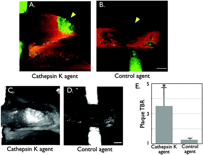
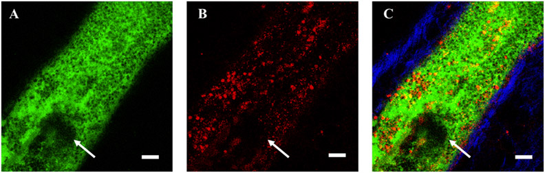
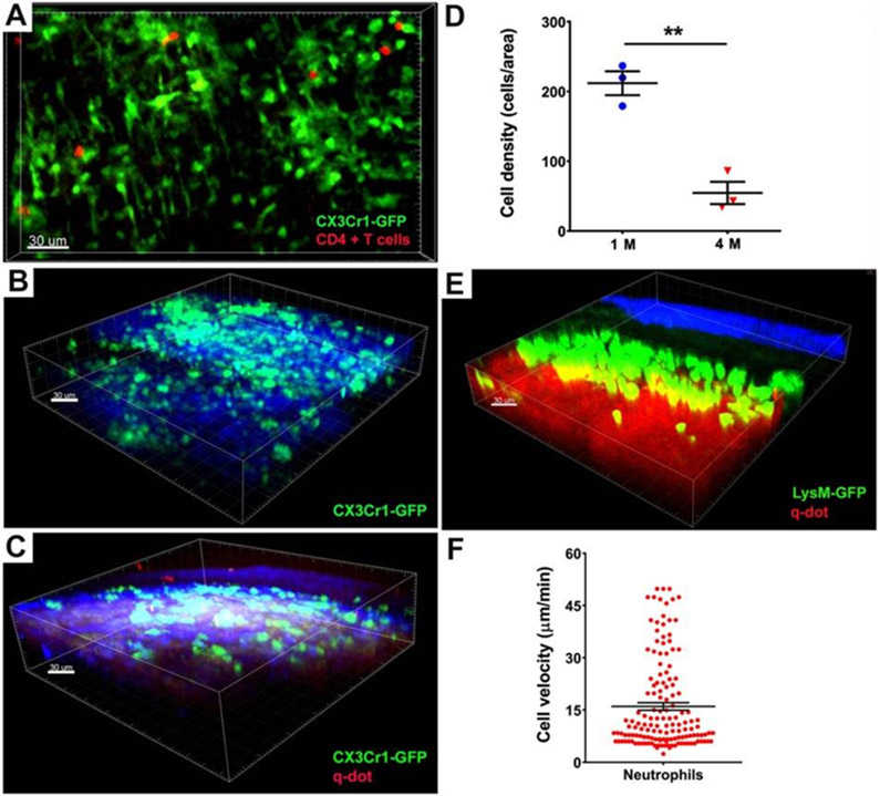
Atherosclerosis is a lipid-driven inflammatory disorder that narrows the arterial lumen and can induce life-threatening complications from coronary artery disease, cerebrovascular disease, and peripheral artery disease. On a mechanistic level, the development of novel cellular-resolution intravital microscopy imaging approaches has recently enabled in vivo studies of underlying biological processes governing disease onset and progress. In particular, multiphoton microscopy has emerged as a promising intravital imaging tool utilizing two-photon-excited fluorescence and second-harmonic generation that provides subcellular resolution and increased imaging depths beyond confocal and epifluorescence microscopy. In this chapter, we describe the state-of-the-art multiphoton microscopy applied to the study of murine atherosclerosis.
动脉粥样硬化是一种由脂质驱动的炎症性疾病,可导致冠状动脉疾病、脑血管疾病和外周动脉疾病等危及生命的并发症。在机制层面上,新型细胞分辨率活体显微镜成像方法的发展最近使得能够对控制疾病发生和进展的潜在生物学过程进行体内研究。特别是,多光子显微镜已成为一种很有前途的活体成像工具,利用双光子激发荧光和二次谐波产生,提供亚细胞分辨率和超过共聚焦和明场显微镜的更深的成像深度。在本章中,我们描述了应用于研究小鼠动脉粥样硬化的最先进的多光子显微镜技术。