Department of Physiology and Biomedical Engineering, Mayo Clinic College of Medicine and Science, 200 First Street SW, Rochester, MN, 55905, USA.
Department of Quantitative Health Sciences, Mayo Clinic College of Medicine and Science, Rochester, MN, 55905, USA.
Breast Cancer Res. 2022 Mar 5;24(1):16. doi: 10.1186/s13058-022-01511-5.
Low specificity in current breast imaging modalities leads to increased unnecessary follow-ups and biopsies. The purpose of this study is to evaluate the efficacy of combining the quantitative parameters of high-definition microvasculature imaging (HDMI) and 2D shear wave elastography (SWE) with clinical factors (lesion depth and age) for improving breast lesion differentiation.
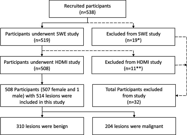
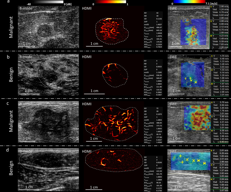
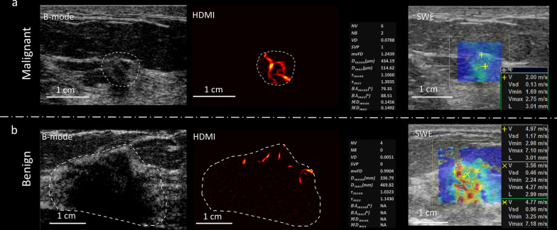
In this prospective study, from June 2016 through April 2021, patients with breast lesions identified on diagnostic ultrasound and recommended for core needle biopsy were recruited. HDMI and SWE were conducted prior to biopsies. Two new HDMI parameters, Murray's deviation and bifurcation angle, and a new SWE parameter, mass characteristic frequency, were included for quantitative analysis. Lesion malignancy prediction models based on HDMI only, SWE only, the combination of HDMI and SWE, and the combination of HDMI, SWE and clinical factors were trained via elastic net logistic regression with 70% (360/514) randomly selected data and validated with the remaining 30% (154/514) data. Prediction performances in the validation test set were compared across models with respect to area under the ROC curve as well as sensitivity and specificity based on optimized threshold selection.
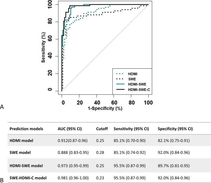
A total of 508 participants (mean age, 54 years ± 15), including 507 female participants and 1 male participant, with 514 suspicious breast lesions (range, 4-72 mm, median size, 13 mm) were included. Of the lesions, 204 were malignant. The SWE-HDMI prediction model, combining quantitative parameters from SWE and HDMI, with AUC of 0.973 (95% CI 0.95-0.99), was significantly higher than the result predicted with the SWE model or HDMI model alone. With an optimal cutoff of 0.25 for the malignancy probability, the sensitivity and specificity were 95.5% and 89.7%, respectively. The specificity was further improved with the addition of clinical factors. The corresponding model defined as the SWE-HDMI-C prediction model had an AUC of 0.981 (95% CI 0.96-1.00).
The SWE-HDMI-C detection model, a combination of SWE estimates, HDMI quantitative biomarkers and clinical factors, greatly improved the accuracy in breast lesion characterization.
当前的乳腺成像方式特异性低,导致不必要的随访和活检增加。本研究旨在评估结合高清微血管成像(HDMI)和二维剪切波弹性成像(SWE)的定量参数与临床因素(病变深度和年龄)对改善乳腺病变鉴别诊断的效果。
本前瞻性研究于 2016 年 6 月至 2021 年 4 月期间,招募了在诊断性超声检查中发现并建议进行核心针活检的乳腺病变患者。在活检前进行 HDMI 和 SWE 检查。纳入了两个新的 HDMI 参数,即默里偏差和分叉角,以及一个新的 SWE 参数,即质量特征频率,用于定量分析。基于 70%(360/514)随机数据,利用弹性网逻辑回归训练了基于 HDMI 仅、SWE 仅、HDMI 和 SWE 组合以及 HDMI、SWE 和临床因素组合的病变恶性预测模型,并利用剩余的 30%(154/514)数据进行验证。在验证测试集中,比较了不同模型的 ROC 曲线下面积以及基于优化阈值选择的敏感性和特异性。
共有 508 名参与者(平均年龄 54 岁±15 岁),包括 507 名女性和 1 名男性,共 514 个可疑乳腺病变(范围 4-72 mm,中位数大小 13 mm)。其中 204 个为恶性病变。SWE-HDMI 预测模型,结合 SWE 和 HDMI 的定量参数,AUC 为 0.973(95%CI 0.95-0.99),明显高于单独使用 SWE 模型或 HDMI 模型的预测结果。当恶性概率的最佳截断值为 0.25 时,敏感性和特异性分别为 95.5%和 89.7%。加入临床因素后,特异性进一步提高。定义为 SWE-HDMI-C 检测模型的相应模型的 AUC 为 0.981(95%CI 0.96-1.00)。
SWE-HDMI-C 检测模型,结合 SWE 估计值、HDMI 定量生物标志物和临床因素,极大地提高了乳腺病变特征的准确性。