Department of Physics and Astronomy, School of Arts and Sciences, Rutgers University, 136 Frelinghuysen Road, Piscataway, NJ, 08854, USA.
Department Physics and Astronomy, Rutgers University, DLS Building, 145 Bevier Road, Room 108, Piscataway, NJ, 08854, USA.
Sci Rep. 2022 Mar 8;12(1):3794. doi: 10.1038/s41598-022-07867-0.
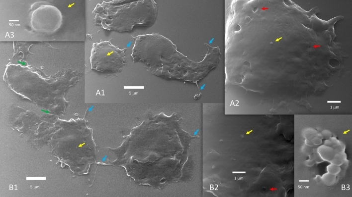
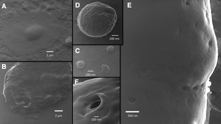
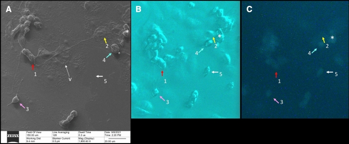
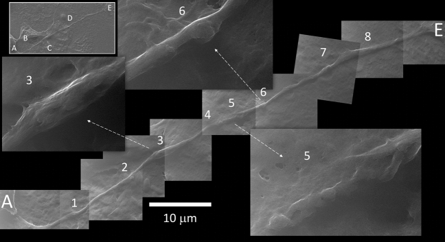
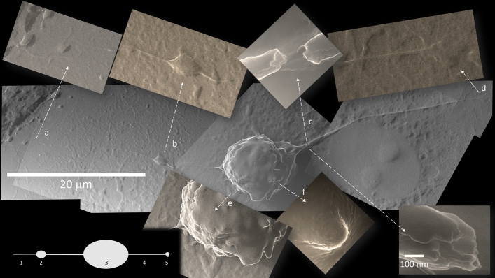
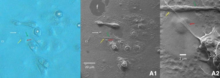
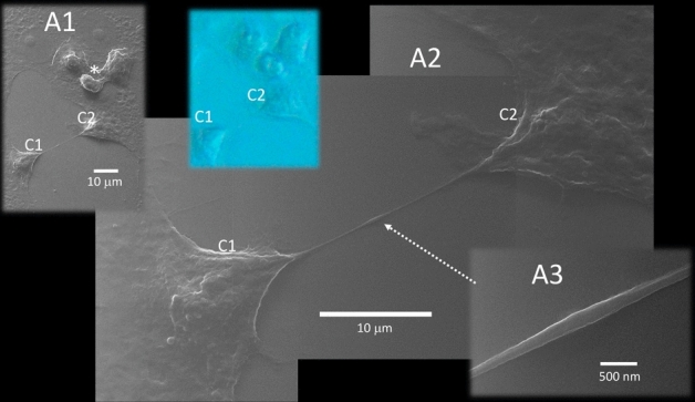
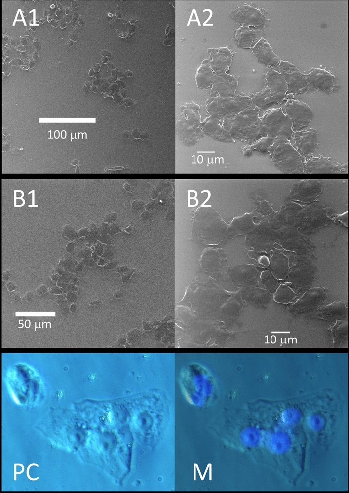
SARS-CoV-2 virions enter the host cells by docking their spike glycoproteins to the membrane-bound Angiotensin Converting Enzyme 2. After intracellular assembly, the newly formed virions are released from the infected cells to propagate the infection, using the extra-cytoplasmic ACE2 docking mechanism. However, the molecular events underpinning SARS-CoV-2 transmission between host cells are not fully understood. Here, we report the findings of a scanning Helium-ion microscopy study performed on Vero E6 cells infected with mNeonGreen-expressing SARS-CoV-2. Our data reveal, with unprecedented resolution, the presence of: (1) long tunneling nanotubes that connect two or more host cells over submillimeter distances; (2) large scale multiple cell fusion events (syncytia); and (3) abundant extracellular vesicles of various sizes. Taken together, these ultrastructural features describe a novel intra-cytoplasmic connection among SARS-CoV-2 infected cells that may act as an alternative route of viral transmission, disengaged from the well-known extra-cytoplasmic ACE2 docking mechanism. Such route may explain the elusiveness of SARS-CoV-2 to survive from the immune surveillance of the infected host.
SARS-CoV-2 病毒粒子通过将其刺突糖蛋白与膜结合的血管紧张素转换酶 2 对接进入宿主细胞。在细胞内组装后,新形成的病毒粒子通过细胞外 ACE2 对接机制从感染的细胞中释放出来以传播感染。然而,宿主细胞之间 SARS-CoV-2 传播的基础分子事件尚不完全清楚。在这里,我们报告了对用 mNeonGreen 表达的 SARS-CoV-2 感染的 Vero E6 细胞进行扫描氦离子显微镜研究的结果。我们的数据以前所未有的分辨率揭示了以下存在:(1)长隧道纳米管,它们在亚毫米距离上连接两个或更多宿主细胞;(2)大规模的多个细胞融合事件(合胞体);和(3)各种大小的丰富细胞外囊泡。这些超微结构特征共同描述了 SARS-CoV-2 感染细胞之间的一种新的细胞内连接,它可能作为一种替代的病毒传播途径,与众所周知的细胞外 ACE2 对接机制无关。这种途径可以解释 SARS-CoV-2 如何逃避感染宿主的免疫监视。