• 文献检索
  • 文档翻译
  • 深度研究
  • 学术资讯
  • Suppr Zotero 插件Zotero 插件
  • 邀请有礼
  • 套餐&价格
  • 历史记录
应用&插件
Suppr Zotero 插件Zotero 插件浏览器插件Mac 客户端Windows 客户端微信小程序
定价
高级版会员购买积分包购买API积分包
服务
文献检索文档翻译深度研究API 文档MCP 服务
关于我们
关于 Suppr公司介绍联系我们用户协议隐私条款
关注我们

Suppr 超能文献

核心技术专利:CN118964589B侵权必究
粤ICP备2023148730 号-1Suppr @ 2026

文献检索

告别复杂PubMed语法,用中文像聊天一样搜索,搜遍4000万医学文献。AI智能推荐,让科研检索更轻松。

立即免费搜索

文件翻译

保留排版,准确专业,支持PDF/Word/PPT等文件格式,支持 12+语言互译。

免费翻译文档

深度研究

AI帮你快速写综述,25分钟生成高质量综述,智能提取关键信息,辅助科研写作。

立即免费体验

Vegfc-Emilin2a-Cxcl8a 信号轴对于斑马鱼心脏再生是必需的。

A Vegfc-Emilin2a-Cxcl8a Signaling Axis Required for Zebrafish Cardiac Regeneration.

机构信息

Department of Developmental Genetics (H.E.-S., R.M.-J., D.Y.R.S.), Max Planck Institute for Heart and Lung Research, Bad Nauheim, Germany.

German Centre for Cardiovascular Research (DZHK) Partner Site Rhine-Main (H.E.-S., S.G., D.Y.R.S.), Max Planck Institute for Heart and Lung Research, Bad Nauheim, Germany.

出版信息

Circ Res. 2022 Apr;130(7):1014-1029. doi: 10.1161/CIRCRESAHA.121.319929. Epub 2022 Mar 10.

DOI:10.1161/CIRCRESAHA.121.319929
PMID:35264012
原文链接:https://pmc.ncbi.nlm.nih.gov/articles/PMC8976759/
Abstract

BACKGROUND

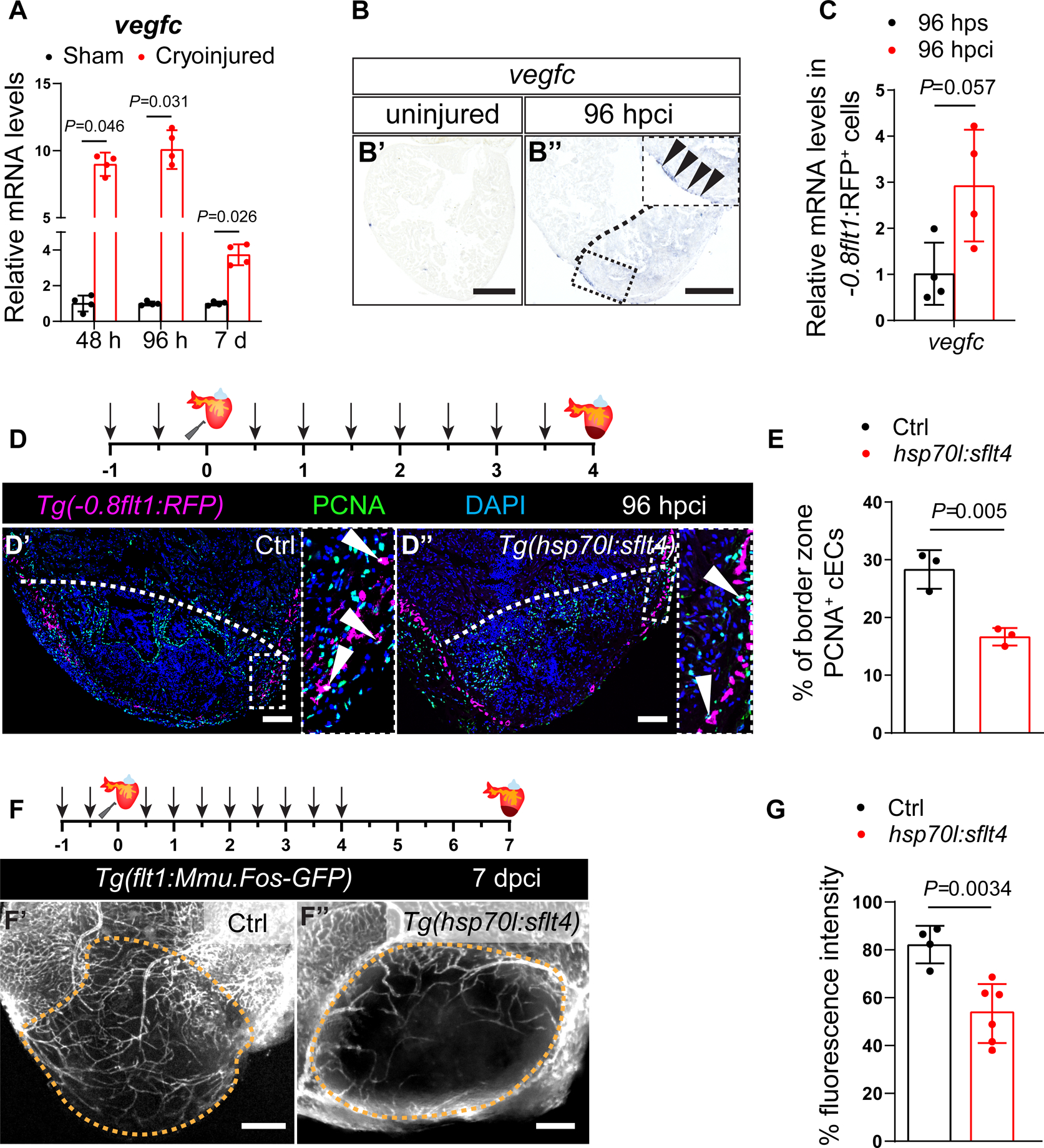
Ischemic heart disease following the obstruction of coronary vessels leads to the death of cardiac tissue and the formation of a fibrotic scar. In contrast to adult mammals, zebrafish can regenerate their heart after injury, enabling the study of the underlying mechanisms. One of the earliest responses following cardiac injury in adult zebrafish is coronary revascularization. Defects in this process lead to impaired cardiomyocyte repopulation and scarring. Hence, identifying and investigating factors that promote coronary revascularization holds great therapeutic potential.

METHODS

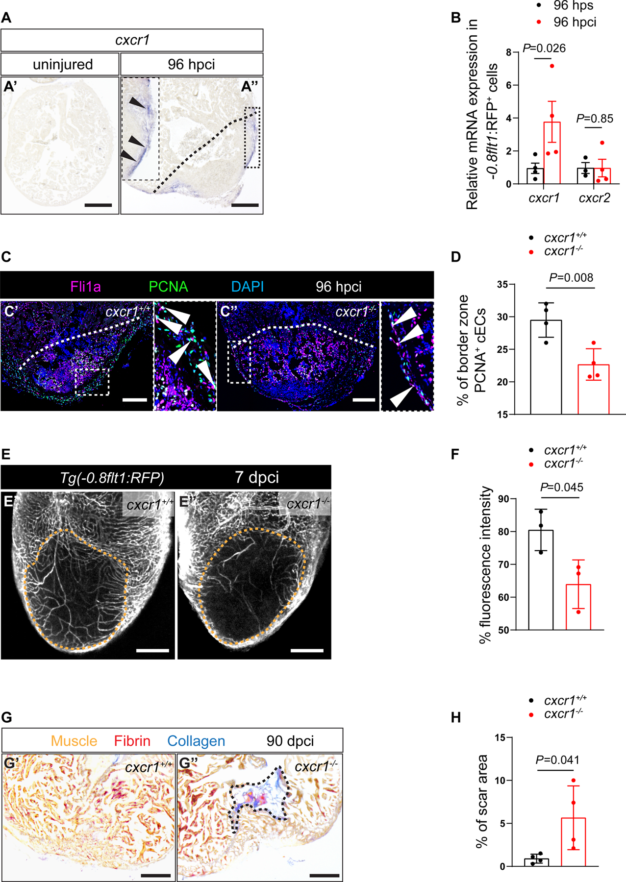
We used wholemount imaging, immunohistochemistry and histology to assess various aspects of zebrafish cardiac regeneration. Deep transcriptomic analysis allowed us to identify targets and potential effectors of Vegfc (vascular endothelial growth factor C) signaling. We used newly generated loss- and gain-of-function genetic tools to investigate the role of Emilin2a (elastin microfibril interfacer 2a) and Cxcl8a (chemokine (C-X-C) motif ligand 8a)-Cxcr1 (chemokine (C-X-C) motif receptor 1) signaling in cardiac regeneration.

RESULTS

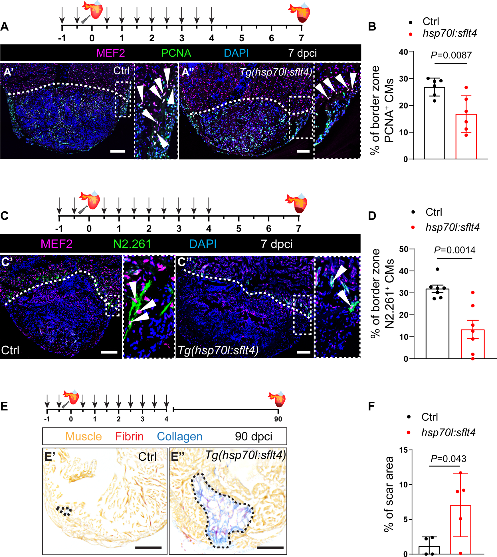
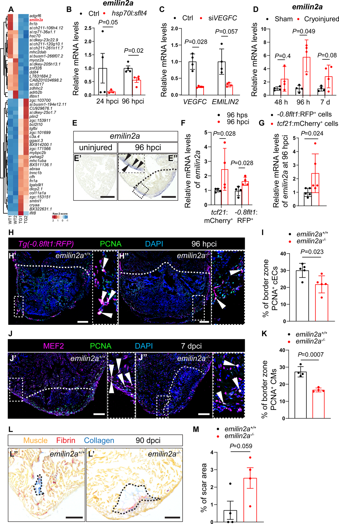
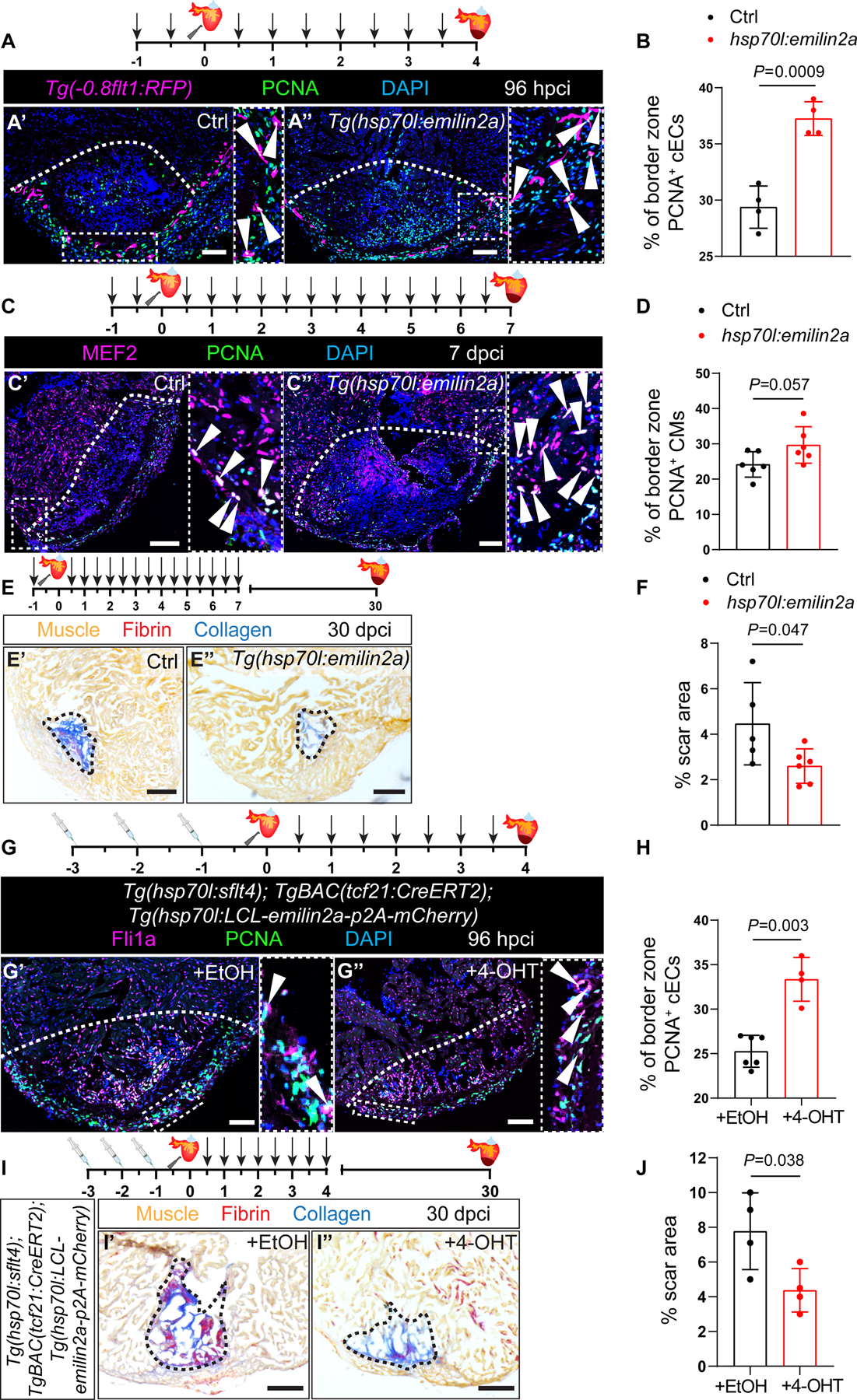
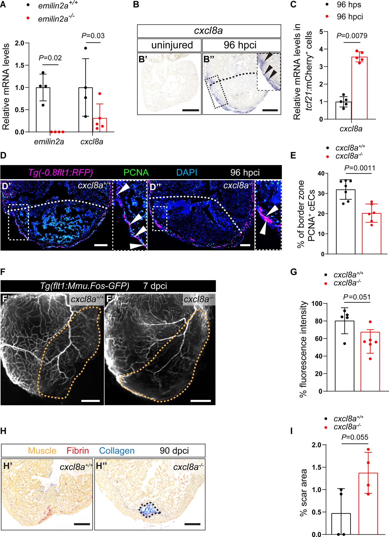
We first show that regenerating coronary endothelial cells upregulate upon cardiac injury in adult zebrafish and that Vegfc signaling is required for their proliferation during regeneration. Notably, blocking Vegfc signaling also significantly reduces cardiomyocyte dedifferentiation and proliferation. Using transcriptomic analyses, we identified as a target of Vegfc signaling and found that manipulation of expression can modulate coronary revascularization as well as cardiomyocyte proliferation. Mechanistically, Emilin2a induces the expression of the chemokine gene in epicardium-derived cells, while , the Cxcl8a receptor gene, is expressed in coronary endothelial cells. We further show that Cxcl8a-Cxcr1 signaling is also required for coronary endothelial cell proliferation during cardiac regeneration.

CONCLUSIONS

These data show that after cardiac injury, coronary endothelial cells upregulate to promote coronary network reestablishment and cardiac regeneration. Mechanistically, Vegfc signaling upregulates epicardial and expression to promote cardiac regeneration. These studies aid in understanding the mechanisms underlying coronary revascularization in zebrafish, with potential therapeutic implications to enhance revascularization and regeneration in injured human hearts.

摘要

背景

冠状动脉阻塞导致心肌组织死亡和形成纤维疤痕,从而引发缺血性心脏病。与成年哺乳动物不同,斑马鱼受伤后可以再生心脏,这使其成为研究潜在机制的理想模型。成年斑马鱼心脏损伤后的最早反应之一是冠状动脉再血管化。该过程中的缺陷会导致心肌细胞再增殖和疤痕形成受损。因此,鉴定和研究促进冠状动脉再血管化的因素具有很大的治疗潜力。

方法

我们使用全胚胎成像、免疫组织化学和组织学来评估斑马鱼心脏再生的各个方面。深度转录组分析使我们能够鉴定 Vegfc(血管内皮生长因子 C)信号的靶标和潜在效应物。我们使用新生成的基因敲除和过表达遗传工具来研究 Emilin2a(弹性微纤维界面蛋白 2a)和 Cxcl8a(趋化因子(C-X-C)基序配体 8a)-Cxcr1(趋化因子(C-X-C)基序受体 1)信号在心脏再生中的作用。

结果

我们首先表明,再生的冠状动脉内皮细胞在成年斑马鱼的心脏损伤后上调 ,并且 Vegfc 信号对于它们在再生过程中的增殖是必需的。值得注意的是,阻断 Vegfc 信号也显著减少了心肌细胞去分化和增殖。通过转录组分析,我们鉴定出 作为 Vegfc 信号的靶标,并发现 表达的操纵可以调节冠状动脉再血管化以及心肌细胞增殖。在机制上,Emilin2a 诱导心外膜衍生细胞中趋化因子基因 的表达,而 ,即 Cxcl8a 受体基因,则在冠状动脉内皮细胞中表达。我们进一步表明,Cxcl8a-Cxcr1 信号在心脏再生过程中也需要冠状动脉内皮细胞增殖。

结论

这些数据表明,心脏损伤后,冠状动脉内皮细胞上调 以促进冠状动脉网络重建和心脏再生。在机制上,Vegfc 信号上调心外膜 和 的表达以促进心脏再生。这些研究有助于理解斑马鱼冠状动脉再血管化的机制,具有增强受损人心肌再血管化和再生的潜在治疗意义。

https://cdn.ncbi.nlm.nih.gov/pmc/blobs/00a2/8976759/4d0a78d52625/nihms-1786113-f0007.jpg
https://cdn.ncbi.nlm.nih.gov/pmc/blobs/00a2/8976759/a7a166fff02f/nihms-1786113-f0002.jpg
https://cdn.ncbi.nlm.nih.gov/pmc/blobs/00a2/8976759/eb77f1030fa4/nihms-1786113-f0003.jpg
https://cdn.ncbi.nlm.nih.gov/pmc/blobs/00a2/8976759/edeec0421adb/nihms-1786113-f0004.jpg
https://cdn.ncbi.nlm.nih.gov/pmc/blobs/00a2/8976759/e256636a6fc6/nihms-1786113-f0005.jpg
https://cdn.ncbi.nlm.nih.gov/pmc/blobs/00a2/8976759/9970fc0a9960/nihms-1786113-f0006.jpg
https://cdn.ncbi.nlm.nih.gov/pmc/blobs/00a2/8976759/4d0a78d52625/nihms-1786113-f0007.jpg

相似文献

1
A Vegfc-Emilin2a-Cxcl8a Signaling Axis Required for Zebrafish Cardiac Regeneration.Vegfc-Emilin2a-Cxcl8a 信号轴对于斑马鱼心脏再生是必需的。
Circ Res. 2022 Apr;130(7):1014-1029. doi: 10.1161/CIRCRESAHA.121.319929. Epub 2022 Mar 10.
2
Coronary Revascularization During Heart Regeneration Is Regulated by Epicardial and Endocardial Cues and Forms a Scaffold for Cardiomyocyte Repopulation.心脏再生过程中心脏冠状动脉的重建受心外膜和心内膜信号的调控,并为心肌细胞再灌注形成支架。
Dev Cell. 2019 Nov 18;51(4):503-515.e4. doi: 10.1016/j.devcel.2019.10.019.
3
Fast revascularization of the injured area is essential to support zebrafish heart regeneration.受伤区域的快速血管再生对于支持斑马鱼心脏再生至关重要。
Proc Natl Acad Sci U S A. 2016 Oct 4;113(40):11237-11242. doi: 10.1073/pnas.1605431113. Epub 2016 Sep 19.
4
Late developing cardiac lymphatic vasculature supports adult zebrafish heart function and regeneration.晚期发育的心脏淋巴管脉管系统支持成年斑马鱼的心脏功能和再生。
Elife. 2019 Nov 8;8:e42762. doi: 10.7554/eLife.42762.
5
Macrophages trigger cardiomyocyte proliferation by increasing epicardial vegfaa expression during larval zebrafish heart regeneration.巨噬细胞通过在斑马鱼幼虫心脏再生过程中增加心外膜 vegfaa 的表达来触发心肌细胞增殖。
Dev Cell. 2022 Jun 20;57(12):1512-1528.e5. doi: 10.1016/j.devcel.2022.05.014. Epub 2022 Jun 9.
6
Zebrafish heart regeneration after coronary dysfunction-induced cardiac damage.冠状动脉功能障碍诱导的心脏损伤后斑马鱼心脏的再生
Dev Biol. 2022 Jul;487:57-66. doi: 10.1016/j.ydbio.2022.04.008. Epub 2022 Apr 29.
7
Primary contribution to zebrafish heart regeneration by gata4(+) cardiomyocytes.Gata4(+) 心肌细胞对斑马鱼心脏再生的主要贡献。
Nature. 2010 Mar 25;464(7288):601-5. doi: 10.1038/nature08804.
8
Defines an Epicardial Cell Subpopulation Required for Cardiomyocyte Expansion During Heart Morphogenesis and Regeneration.定义了在心脏形态发生和再生过程中心肌细胞扩增所需的心外膜细胞亚群。
Circulation. 2022 Jul 5;146(1):48-63. doi: 10.1161/CIRCULATIONAHA.121.055468. Epub 2022 Jun 2.
9
Migration of cardiomyocytes is essential for heart regeneration in zebrafish.心肌细胞的迁移对于斑马鱼的心脏再生至关重要。
Development. 2012 Nov;139(22):4133-42. doi: 10.1242/dev.079756. Epub 2012 Oct 3.
10
Igf Signaling is Required for Cardiomyocyte Proliferation during Zebrafish Heart Development and Regeneration.胰岛素样生长因子信号传导在斑马鱼心脏发育和再生过程中对心肌细胞增殖是必需的。
PLoS One. 2013 Jun 26;8(6):e67266. doi: 10.1371/journal.pone.0067266. Print 2013.

引用本文的文献

1
Efficacy and safety of empagliflozin in patients over 65 with type 2 diabetes mellitus complicating cardiorenal syndromes type II and IV.恩格列净在65岁以上2型糖尿病合并II型和IV型心肾综合征患者中的疗效和安全性。
J Med Biochem. 2025 Jul 4;44(4):905-914. doi: 10.5937/jomb0-54743.
2
Macrophages and cardiac lesion in zebrafish: what can single-cell RNA sequencing reveal?斑马鱼中的巨噬细胞与心脏病变:单细胞RNA测序能揭示什么?
Front Cardiovasc Med. 2025 Apr 11;12:1570582. doi: 10.3389/fcvm.2025.1570582. eCollection 2025.
3
Cardiac Regeneration in Adult Zebrafish: A Review of Signaling and Metabolic Coordination.

本文引用的文献

1
VEGFC/FLT4-induced cell-cycle arrest mediates sprouting and differentiation of venous and lymphatic endothelial cells.VEGFC/FLT4 诱导的细胞周期停滞介导静脉和淋巴管内皮细胞的出芽和分化。
Cell Rep. 2021 Jun 15;35(11):109255. doi: 10.1016/j.celrep.2021.109255.
2
Cardiomyocyte heterogeneity during zebrafish development and regeneration.斑马鱼发育和再生过程中心肌细胞的异质性。
Dev Biol. 2021 Aug;476:259-271. doi: 10.1016/j.ydbio.2021.03.014. Epub 2021 Apr 12.
3
Krüppel-like factor 1 is a core cardiomyogenic trigger in zebrafish.
成年斑马鱼的心脏再生:信号传导与代谢协调综述
Curr Cardiol Rep. 2025 Jan 10;27(1):15. doi: 10.1007/s11886-024-02162-y.
4
flt1 inactivation promotes zebrafish cardiac regeneration by enhancing endothelial activity and limiting the fibrotic response.Flt1 失活通过增强内皮细胞活性和限制纤维化反应促进斑马鱼心脏再生。
Development. 2024 Dec 1;151(23). doi: 10.1242/dev.203028. Epub 2024 Nov 29.
5
Harnessing the regenerative potential of interleukin11 to enhance heart repair.利用白细胞介素 11 的再生潜力增强心脏修复。
Nat Commun. 2024 Nov 8;15(1):9666. doi: 10.1038/s41467-024-54060-0.
6
Differential vegfc expression dictates lymphatic response during zebrafish heart development and regeneration.差异表达的 vegfc 决定斑马鱼心脏发育和再生过程中的淋巴管反应。
Development. 2024 Nov 15;151(22). doi: 10.1242/dev.202947. Epub 2024 Nov 19.
7
The innate immune regulator MyD88 dampens fibrosis during zebrafish heart regeneration.先天免疫调节剂 MyD88 在斑马鱼心脏再生过程中抑制纤维化。
Nat Cardiovasc Res. 2024 Sep;3(9):1158-1176. doi: 10.1038/s44161-024-00538-5. Epub 2024 Sep 13.
8
Exploring the Function of Epicardial Cells Beyond the Surface.探索心外膜细胞的表面下功能。
Circ Res. 2024 Jul 5;135(2):353-371. doi: 10.1161/CIRCRESAHA.124.321567. Epub 2024 Jul 4.
9
Endothelial progenitor cell-derived extracellular vesicles: the world of potential prospects for the treatment of cardiovascular diseases.内皮祖细胞衍生的细胞外囊泡:心血管疾病治疗的潜在前景世界。
Cell Biosci. 2024 Jun 5;14(1):72. doi: 10.1186/s13578-024-01255-z.
10
Antigen presentation plays positive roles in the regenerative response to cardiac injury in zebrafish.抗原呈递在斑马鱼心脏损伤的再生反应中发挥积极作用。
Nat Commun. 2024 Apr 29;15(1):3637. doi: 10.1038/s41467-024-47430-1.
Krüppel-like factor 1 是斑马鱼心脏发生的核心触发因子。
Science. 2021 Apr 9;372(6538):201-205. doi: 10.1126/science.abe2762.
4
Deregulated expression of Elastin Microfibril Interfacer 2 (EMILIN2) in gastric cancer affects tumor growth and angiogenesis.弹性微原纤维界面蛋白2(EMILIN2)在胃癌中的异常表达影响肿瘤生长和血管生成。
Matrix Biol Plus. 2020 Feb 19;6-7:100029. doi: 10.1016/j.mbplus.2020.100029. eCollection 2020 May.
5
Modulation of Mammalian Cardiomyocyte Cytokinesis by the Extracellular Matrix.细胞外基质对哺乳动物心肌细胞胞质分裂的调节
Circ Res. 2020 Sep 11;127(7):896-907. doi: 10.1161/CIRCRESAHA.119.316303. Epub 2020 Jun 21.
6
Runx1 promotes scar deposition and inhibits myocardial proliferation and survival during zebrafish heart regeneration.Runx1 在斑马鱼心脏再生过程中促进疤痕沉积,并抑制心肌增殖和存活。
Development. 2020 Apr 27;147(8):dev186569. doi: 10.1242/dev.186569.
7
AP-1 Contributes to Chromatin Accessibility to Promote Sarcomere Disassembly and Cardiomyocyte Protrusion During Zebrafish Heart Regeneration.AP-1 促进染色质可及性以促进斑马鱼心脏再生过程中的肌节解体和心肌细胞突起。
Circ Res. 2020 Jun 5;126(12):1760-1778. doi: 10.1161/CIRCRESAHA.119.316167. Epub 2020 Apr 21.
8
Genetic lineage tracing reveals poor angiogenic potential of cardiac endothelial cells.遗传谱系追踪揭示了心脏内皮细胞的不良血管生成潜力。
Cardiovasc Res. 2021 Jan 1;117(1):256-270. doi: 10.1093/cvr/cvaa012.
9
Coronary Revascularization During Heart Regeneration Is Regulated by Epicardial and Endocardial Cues and Forms a Scaffold for Cardiomyocyte Repopulation.心脏再生过程中心脏冠状动脉的重建受心外膜和心内膜信号的调控,并为心肌细胞再灌注形成支架。
Dev Cell. 2019 Nov 18;51(4):503-515.e4. doi: 10.1016/j.devcel.2019.10.019.
10
Distinct origins and molecular mechanisms contribute to lymphatic formation during cardiac growth and regeneration.在心脏生长和再生过程中,淋巴管的形成有不同的起源和分子机制。
Elife. 2019 Nov 8;8:e44153. doi: 10.7554/eLife.44153.