Department of Orthopedics, Orthopedic Research Institute, West China Hospital, Sichuan University, No. 37 Guoxuexiang, Chengdu, 610041, Sichuan, People's Republic of China.
J Orthop Surg Res. 2022 Mar 9;17(1):151. doi: 10.1186/s13018-022-03039-y.
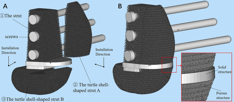
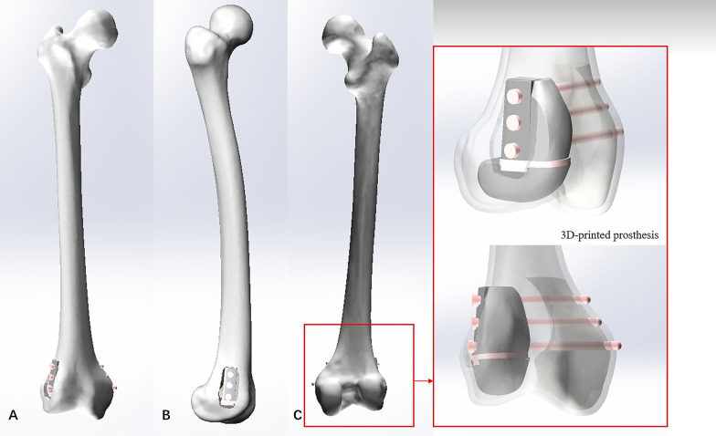
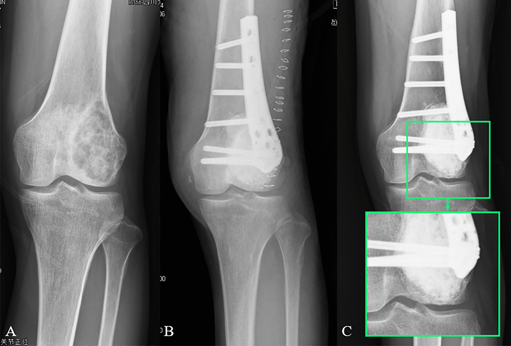
The most common reconstruction method for bone defects caused by giant cell tumor of bone (GCTB) is cement packing combined with subchondral bone grafting and extra fixation. However, this method has several limitations involving bone cement and bone graft, which may lead to poor prognosis and joint function. A titanium-based 3D-printed strut-type prosthesis, featured with excellent biocompatibility and osseointegration ability, was developed for this bone defect in our institution. The goal of this study is to comparatively analyze the biomechanical performance of reconstruction methods aimed at the identification of better operative strategy.
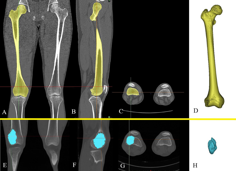
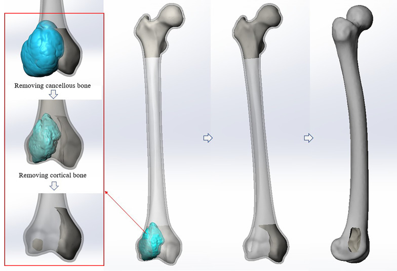
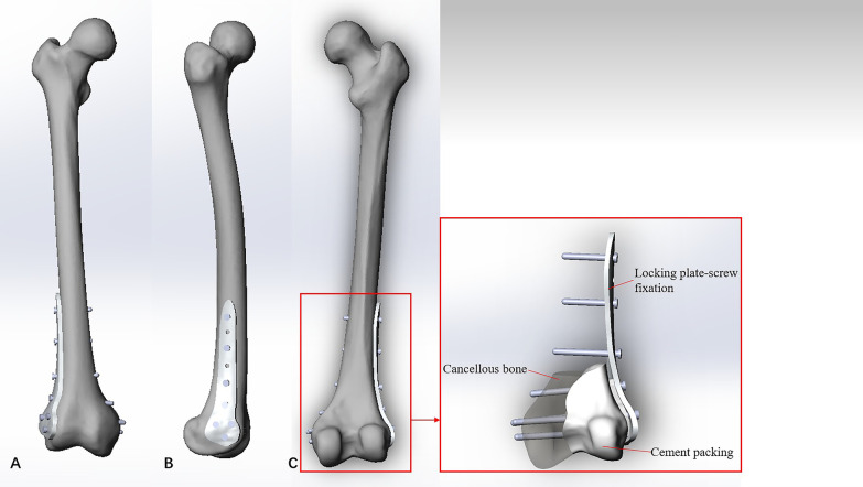
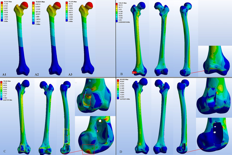
Four different 3D finite element models were created. Model #1: Normal femur; Model #2: Femur with tumorous cavity bone defects in the distal femur; Model #3: Cavity bone defects reconstructed by cement packing combined with subchondral bone grafting and extra fixation; Model #4: Cavity bone defects reconstructed by 3D-printed strut-type prosthesis combined with subchondral bone grafting. The femoral muscle multiple forces were applied to analyze the mechanical difference among these models by finite element analysis.
Optimal stress and displacement distribution were observed in the normal femur. Both reconstruction methods could provide good initial stability and mechanical support. Stress distributed unevenly on the femur repaired by cement packing combined with subchondral bone grafting and extra fixation, and obvious stress concentration was found around the articular surface of this femur. However, the femur repaired by 3D-printed strut-type prosthetic reconstruction showed better performance both in displacement and stress distribution, particularly in terms of the protection of articular surface and subchondral bone.
3D-printed strut-type prosthesis is outstanding in precise shape matching and better osseointegration. Compared to cement packing and extra fixation, it can provide the almost same support and fixation stiffness, but better biomechanical performance and protection of subchondral bone and articular cartilage. Therefore, 3D-printed strut-type prosthetic reconstruction combined with subchondral bone grafting may be evaluated as an alternative for the treatment of GCTBs in distal femur.
骨巨细胞瘤(GCTB)导致的骨缺损最常见的重建方法是骨水泥填充联合软骨下骨移植和额外固定。然而,这种方法存在涉及骨水泥和骨移植物的几个局限性,可能导致预后不良和关节功能不佳。我们机构为这种骨缺损开发了一种基于钛的 3D 打印支撑型假体,具有出色的生物相容性和骨整合能力。本研究的目的是比较分析针对识别更好手术策略的重建方法的生物力学性能。
创建了四个不同的 3D 有限元模型。模型#1:正常股骨;模型#2:股骨远端有肿瘤性腔骨缺损;模型#3:骨水泥填充联合软骨下骨移植和额外固定重建的腔骨缺损;模型#4:3D 打印支撑型假体联合软骨下骨移植重建的腔骨缺损。通过有限元分析,应用股骨肌肉多力来分析这些模型之间的力学差异。
在正常股骨中观察到最佳的应力和位移分布。两种重建方法都可以提供良好的初始稳定性和机械支撑。骨水泥填充联合软骨下骨移植和额外固定重建的股骨应力分布不均匀,股骨关节面附近发现明显的应力集中。然而,3D 打印支撑型假体重建的股骨在位移和应力分布方面表现出更好的性能,特别是在保护关节面和软骨下骨方面。
3D 打印支撑型假体在精确的形状匹配和更好的骨整合方面表现出色。与骨水泥填充和额外固定相比,它可以提供几乎相同的支撑和固定刚度,但具有更好的生物力学性能和对软骨下骨和关节软骨的保护。因此,3D 打印支撑型假体重建联合软骨下骨移植可能被评估为治疗股骨远端 GCTB 的一种替代方法。