Ketschek Andrea, Holland Sabrina M, Gallo Gianluca
Shriners Hospitals Pediatric Research Center, Lewis Katz School of Medicine, Temple University, Philadelphia, PA, United States.
Department of Neural Sciences, Lewis Katz School of Medicine, Temple University, Philadelphia, PA, United States.
Front Mol Neurosci. 2022 Feb 21;15:726962. doi: 10.3389/fnmol.2022.726962. eCollection 2022.
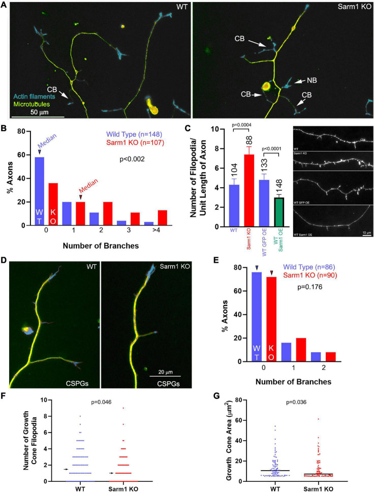
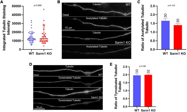
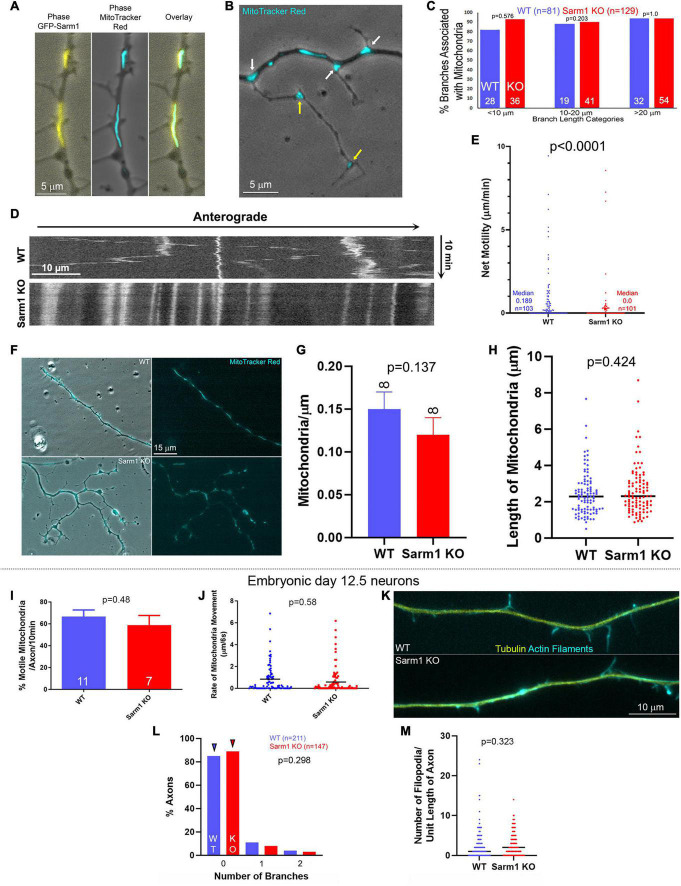
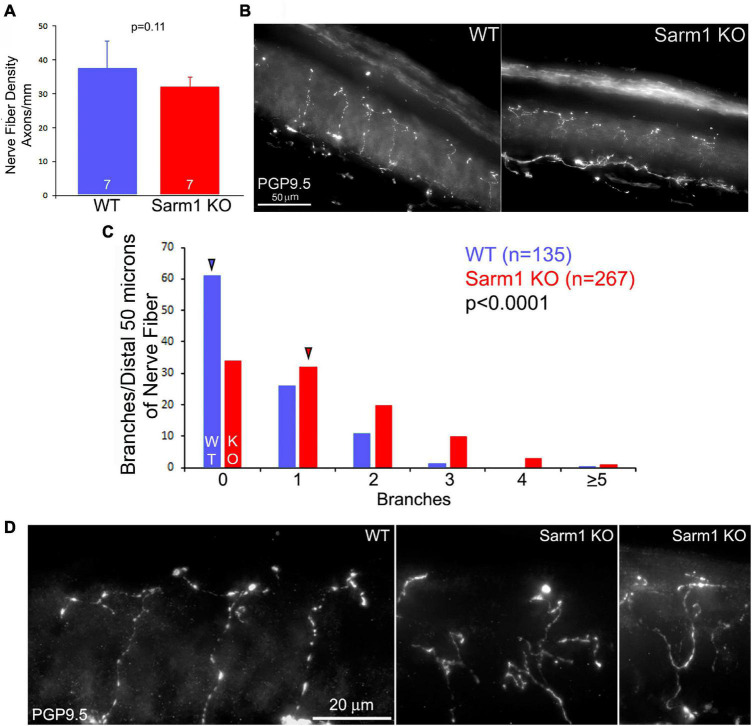
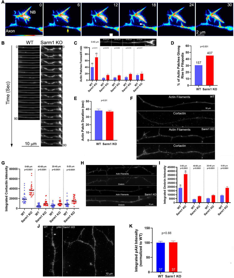
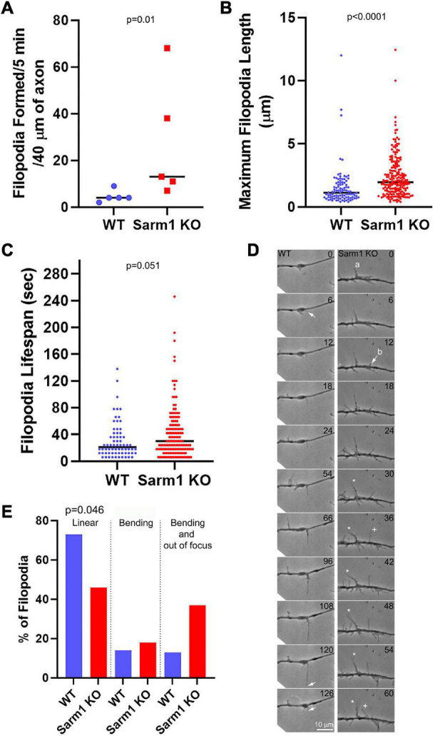
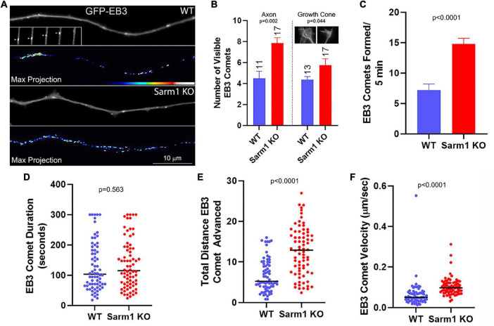
Axon branching is a fundamental aspect of neuronal morphogenesis, neuronal circuit formation, and response of the nervous system to injury. Sterile alpha and TIR motif containing 1 (SARM1) was initially identified as promoting Wallerian degeneration of axons. We now report a novel function of SARM1 in postnatal sensory neurons; the suppression of axon branching. Axon collateral branches develop from axonal filopodia precursors through the coordination of the actin and microtubule cytoskeleton. analysis revealed that cultured P0-2 dorsal root ganglion sensory neurons from a SARM1 knockout (KO) mouse exhibit increased numbers of collateral branches and axonal filopodia relative to wild-type neurons. In SARM1 KO mice, cutaneous sensory endings exhibit increased branching in the skin with normal density of innervation. Transient axonal actin patches serve as cytoskeletal platforms from which axonal filopodia emerge. Live imaging analysis of axonal actin dynamics showed that SARM1 KO neurons exhibit increased rates of axonal actin patch formation and increased probability that individual patches will give rise to a filopodium before dissipating. SARM1 KO axons contain elevated levels of drebrin and cortactin, two actin regulatory proteins that are positive regulators of actin patches, filopodia formation, and branching. Live imaging of microtubule plus tip dynamics revealed an increase in the rate of formation and velocity of polymerizing tips along the axons of SARM1 KO neurons. Stationary mitochondria define sites along the axon where branches may arise, and the axons of SARM1 KO sensory neurons exhibit an increase in stationary mitochondria. These data reveal SARM1 to be a negative regulator of axonal cytoskeletal dynamics and collateral branching.
轴突分支是神经元形态发生、神经回路形成以及神经系统对损伤反应的一个基本方面。含无菌α和TIR基序1(SARM1)最初被确定为促进轴突的华勒氏变性。我们现在报告SARM1在出生后感觉神经元中的一种新功能;抑制轴突分支。轴突侧支从轴突丝状伪足前体通过肌动蛋白和微管细胞骨架的协调发育而来。分析表明,与野生型神经元相比,来自SARM1基因敲除(KO)小鼠的培养的P0 - 2背根神经节感觉神经元表现出侧支和轴突丝状伪足数量增加。在SARM1基因敲除小鼠中,皮肤感觉末梢在皮肤中表现出分支增加且神经支配密度正常。短暂的轴突肌动蛋白斑块作为轴突丝状伪足出现的细胞骨架平台。对轴突肌动蛋白动力学的实时成像分析表明,SARM1基因敲除神经元表现出轴突肌动蛋白斑块形成速率增加,并且单个斑块在消散前产生丝状伪足的概率增加。SARM1基因敲除的轴突含有升高水平的drebrin和cortactin,这两种肌动蛋白调节蛋白是肌动蛋白斑块、丝状伪足形成和分支的确正调节因子。对微管正端动力学的实时成像显示,SARM1基因敲除神经元轴突上聚合末端的形成速率和速度增加。静止的线粒体确定了轴突上可能产生分支的位点,并且SARM1基因敲除感觉神经元的轴突表现出静止线粒体增加。这些数据表明SARM1是轴突细胞骨架动力学和侧支分支的负调节因子。