Yeh Chuan-Feng, Juang Duane S, Chen Ya-Wen, Rodoplu Didem, Hsu Chia-Hsien
Institute of Biomedical Engineering and Nanomedicine, National Health Research Institutes, Miaol, Taiwan.
Institute of NanoEngineering and MicroSystems, National Tsing Hua University, Hsinchu, Taiwan.
Front Bioeng Biotechnol. 2022 Feb 24;10:852318. doi: 10.3389/fbioe.2022.852318. eCollection 2022.
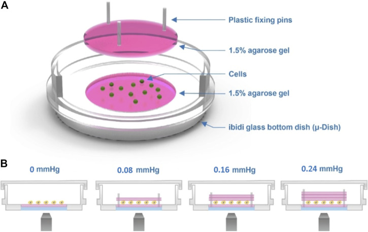
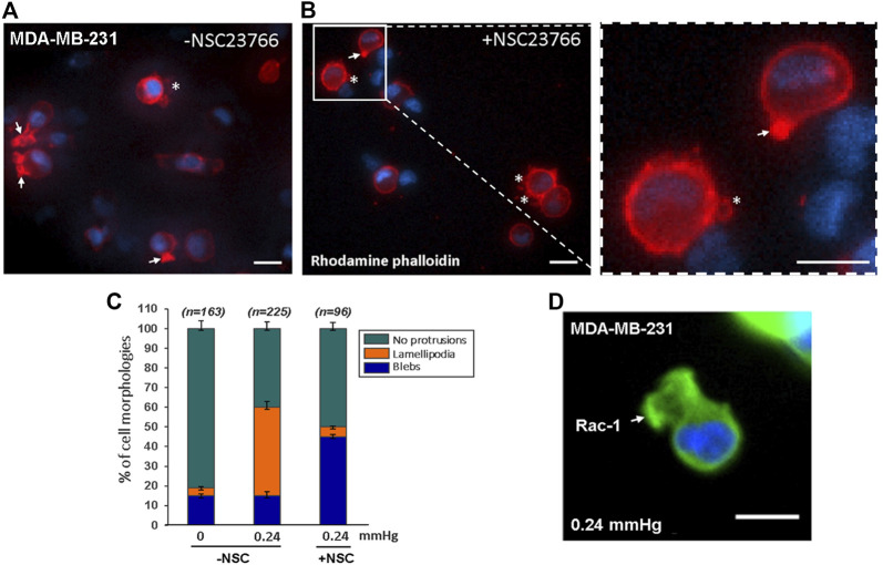
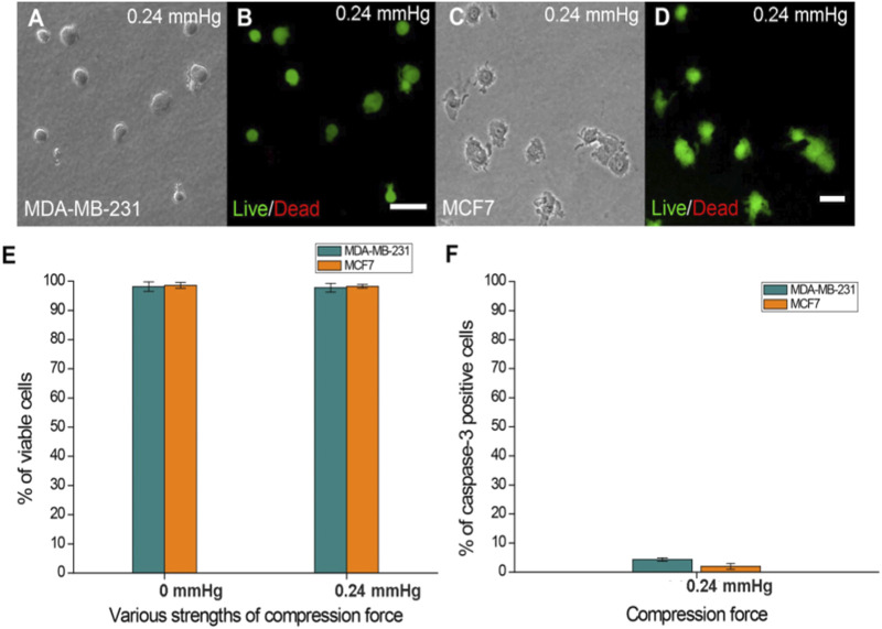
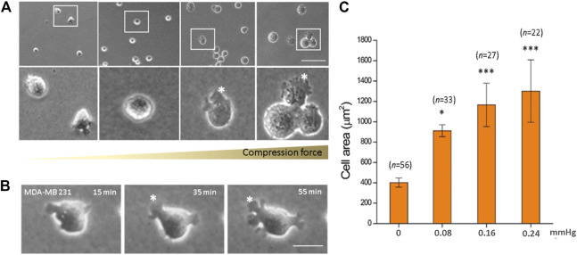
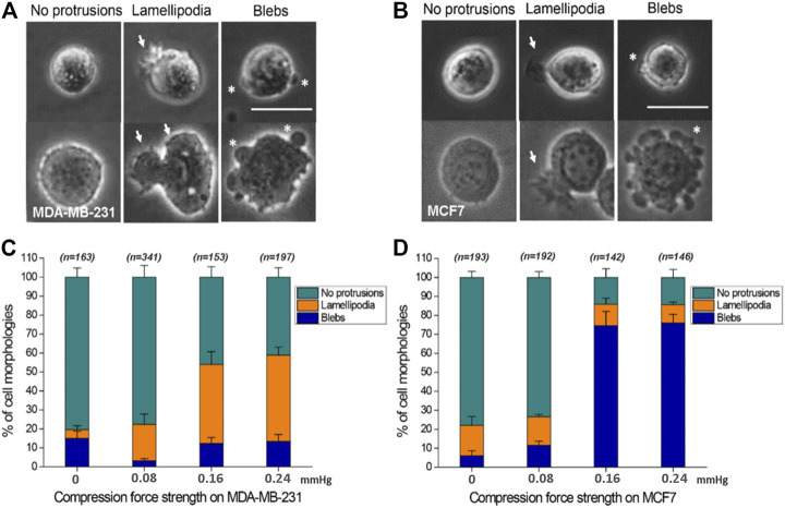
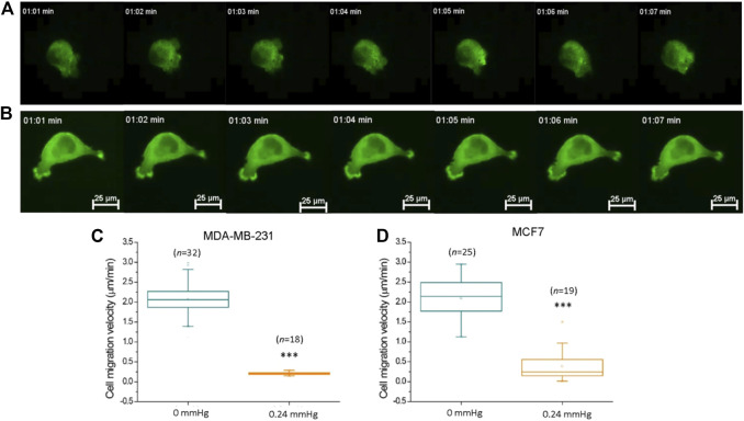
devices offer more numerous methods than models to investigate how cells respond to pressure stress and quantify those responses. Several devices have been developed to study the cell response to compression force. However, they are unable to observe morphological changes of cells in real-time. There is also a concern about cell damage during the process of harvesting cells from 3D gels. Here we report a device employing transparent, thin gel layers to clamp cells between the interfaces and applied a controllable compression force by stacking multiple layers on the top. In this approach, cells can be monitored for alteration of cellular protrusions, whose diversity has been proven to promote cancer cell dissemination, with single-cell resolution under compression force. Furthermore, p-Rac-1 and rhodamine staining on the device directly to confirm the actin filaments of lamellipodia. The method was able to fulfill real-time live-cell observation at single-cell resolution and can be readily used for versatile cell analysis. MDA-MB-231 and MCF7 breast cancer cells were utilized to demonstrate the utility of the device, and the results showed that the stimuli of compression force induce MDA-MB-231 and MCF7 to form lamellipodia and bleb protrusions, respectively. We envision the device may be used as a tool to explore mechanisms of membrane protrusion transitions and to screen drug candidates for inhibiting cancer cell protrusion plasticity for cancer therapy.
与模型相比,设备提供了更多的方法来研究细胞如何应对压力应激并量化这些反应。已经开发了几种设备来研究细胞对压缩力的反应。然而,它们无法实时观察细胞的形态变化。从3D凝胶中收获细胞的过程中还存在细胞损伤的问题。在此,我们报告一种设备,该设备采用透明薄凝胶层将细胞夹在界面之间,并通过在顶部堆叠多层来施加可控的压缩力。通过这种方法,可以在压缩力下以单细胞分辨率监测细胞突起的变化,细胞突起的多样性已被证明可促进癌细胞扩散。此外,在该设备上直接进行p-Rac-1和罗丹明染色以确认片状伪足的肌动蛋白丝。该方法能够以单细胞分辨率实现实时活细胞观察,并且可容易地用于多种细胞分析。利用MDA-MB-231和MCF7乳腺癌细胞来证明该设备的实用性,结果表明压缩力刺激分别诱导MDA-MB-231和MCF7形成片状伪足和气泡状突起。我们设想该设备可作为一种工具,用于探索膜突起转变的机制,并筛选抑制癌细胞突起可塑性以用于癌症治疗的候选药物。