Department of spine surgery, First Affiliated Hospital of Gannan Medical University, No. 128 Jin Ling Road, 341000, Ganzhou, Jiangxi, China.
Department of basic medicine, Gannan Health Vocational College, No. 12 Rong Jiang Road, 341000, Ganzhou, Jiangxi, China.
BMC Endocr Disord. 2022 Mar 14;22(1):66. doi: 10.1186/s12902-022-00971-2.
Brown tumour is a rare tumour-like lesion of the bone, which is considered as an end-stage lesion of abnormal bone metabolism caused by persistently high parathyroid hormone (PTH) levels. Brown tumour can be found in any part of the skeleton; in some cases, it can occur in multiple bones and can be easily misdiagnosed as a metastatic tumour.
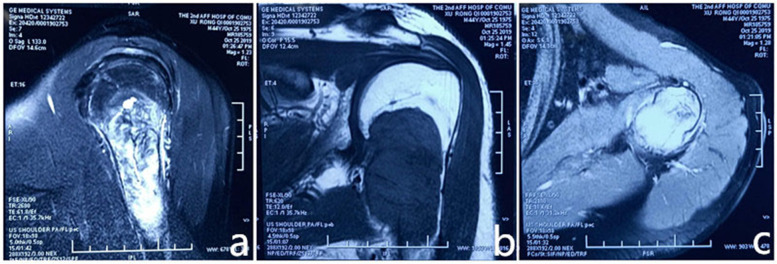
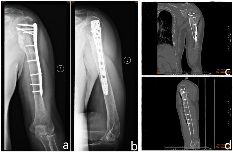
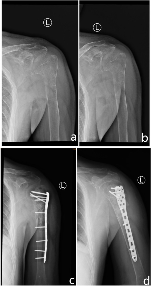
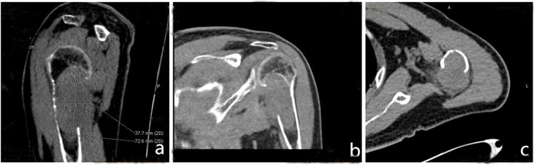
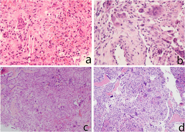
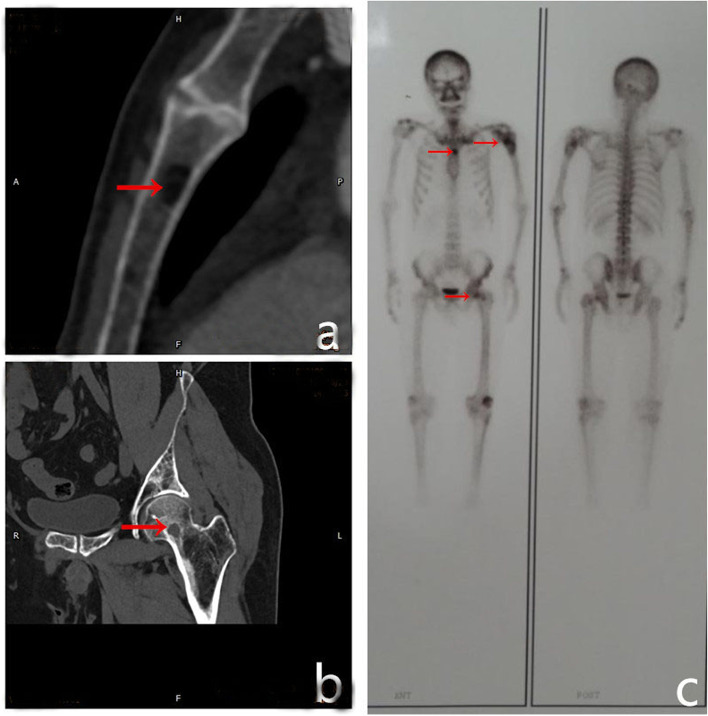
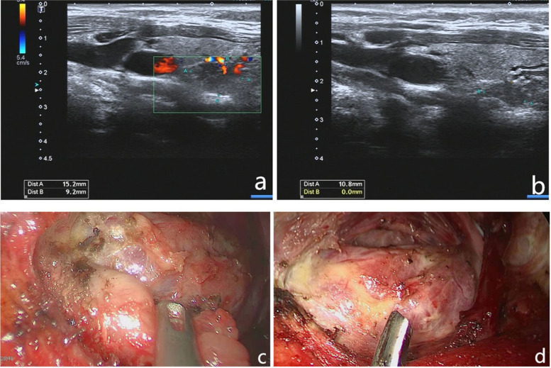
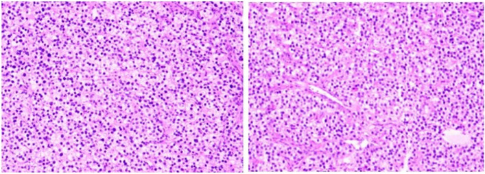
We report the case of a 44-year-old man who presented to the Department of Oncology in our hospital with a 2-month history of local pain in his left shoulder joint. The initial diagnosis was an aneurysmal bone cyst by biopsy, for which the patient underwent tumour resection surgery. The diagnosis of a malignant tumour was made again following postoperative pathological examination. The pathological sections and all clinical data were sent to the Department of Pathology of the First Affiliated Hospital of Sun Yat-sen University; the diagnosis made there was brown tumour. His blood PTH level was 577 pg/ml (15-65 pg/ml). Colour Doppler ultrasonography of the parathyroid gland suggested a parathyroid adenoma. For further treatment, the left parathyroid adenoma was removed by axillary endoscopic resection. Postoperatively, a pathologic examination was performed, and the diagnosis of a parathyroid adenoma was confirmed. One year after the surgery, the left humerus was completely healed, and the left shoulder joint had a good range of movement.
In summary, histopathological diagnosis is not sufficient for the diagnosis of brown tumours. A comprehensive analysis combining clinical symptoms with findings of imaging and laboratory tests is also required. Generally, the treatment of brown tumour includes only partial or complete resection of the parathyroid glands. However, when the tumour is large, especially when it involves the joint, surgery is indispensable.
棕色瘤是一种罕见的骨肿瘤样病变,被认为是由甲状旁腺激素(PTH)水平持续升高引起的异常骨代谢的终末阶段病变。棕色瘤可发生在骨骼的任何部位;在某些情况下,它可以发生在多个骨骼中,并且容易被误诊为转移性肿瘤。
我们报告了一例 44 岁男性患者,因左肩关节局部疼痛 2 个月就诊于我院肿瘤科。最初的诊断是活检提示为动脉瘤样骨囊肿,为此患者接受了肿瘤切除术。术后病理检查再次诊断为恶性肿瘤。术后病理切片及所有临床资料均送至中山大学附属第一医院病理科,诊断为棕色瘤。患者的血 PTH 水平为 577pg/ml(15-65pg/ml)。甲状旁腺的彩色多普勒超声提示甲状旁腺腺瘤。为进一步治疗,采用腋内镜切除左侧甲状旁腺腺瘤。术后行病理检查,诊断为甲状旁腺腺瘤。术后 1 年,左侧肱骨完全愈合,左侧肩关节活动良好。
综上所述,棕色瘤的诊断仅依靠组织病理学检查是不够的,还需要结合临床症状并结合影像学和实验室检查结果进行综合分析。一般来说,棕色瘤的治疗包括仅部分或完全切除甲状旁腺。然而,当肿瘤较大,特别是累及关节时,手术是必不可少的。