Finnish Institute of Occupational Health, Työterveyslaitos, Box 40, 00032, Helsinki, Finland.
Department of Bioproducts and Biosystems, Aalto University, Espoo, Finland.
Part Fibre Toxicol. 2022 Mar 16;19(1):19. doi: 10.1186/s12989-022-00460-3.
Cellulose nanofibrils (CNFs) have emerged as a sustainable and environmentally friendly option for a broad range of applications. The fibrous nature and high biopersistence of CNFs call for a thorough toxicity assessment, but it is presently unclear which physico-chemical properties could play a role in determining the potential toxic response to CNF. Here, we assessed whether surface composition and size could modulate the genotoxicity of CNFs in human bronchial epithelial BEAS-2B cells. We examined three size fractions (fine, medium and coarse) of four CNFs with different surface chemistry: unmodified (U-CNF) and functionalized with 2,2,6,6-tetramethyl-piperidin-1-oxyl (TEMPO) (T-CNF), carboxymethyl (C-CNF) and epoxypropyltrimethylammonium chloride (EPTMAC) (E-CNF). In addition, the source fibre was also evaluated as a non-nanosized material.
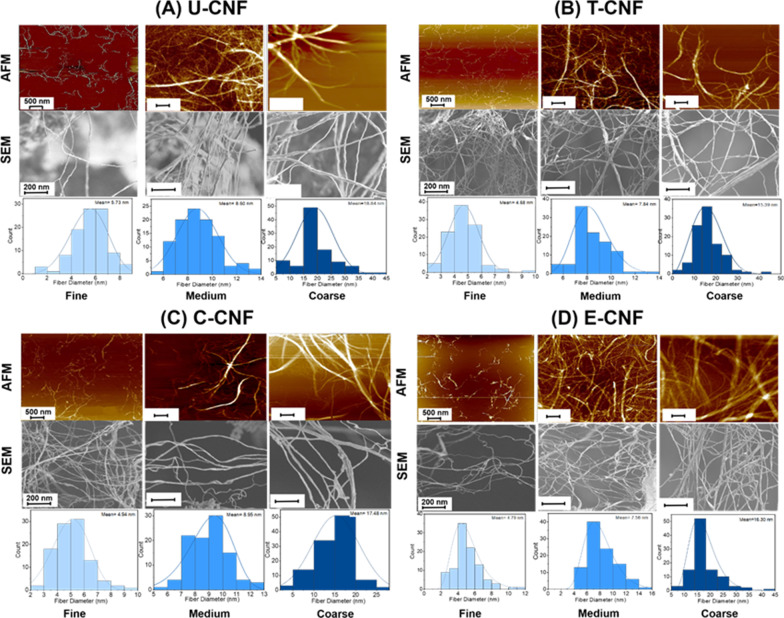
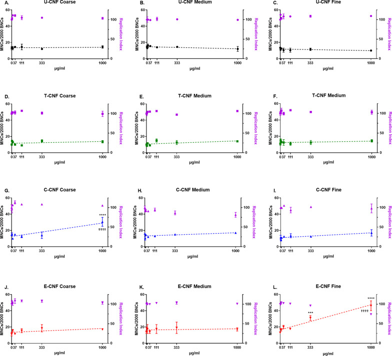
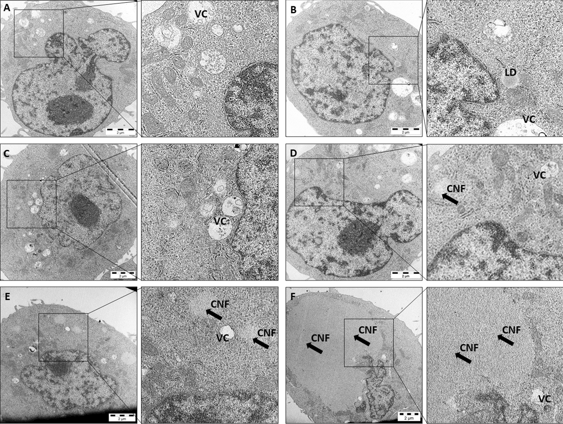
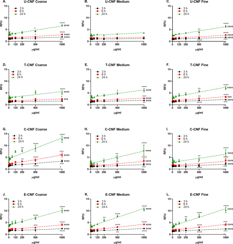
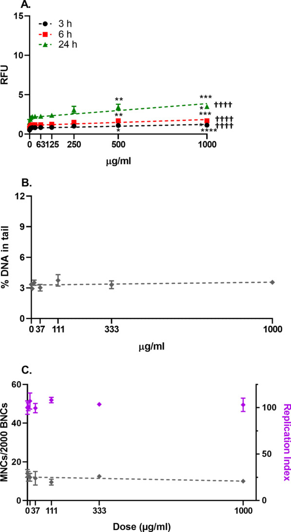
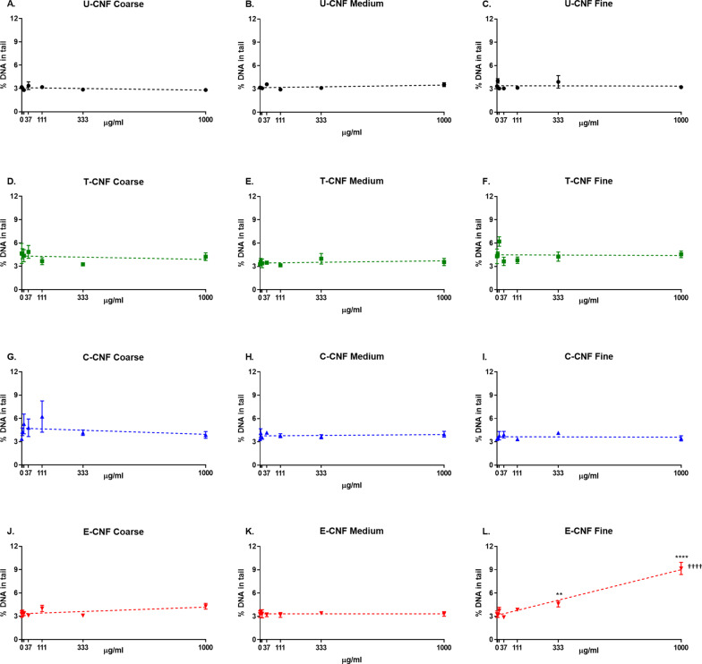
The presence of the surface charged groups in the functionalized CNF samples resulted in higher amounts of individual nanofibrils and less aggregation compared with the U-CNF. T-CNF was the most homogenous, in agreement with its high surface group density. However, the colloidal stability of all the CNF samples dropped when dispersed in cell culture medium, especially in the case of T-CNF. CNF was internalized by a minority of BEAS-2B cells. No remarkable cytotoxic effects were induced by any of the cellulosic materials. All cellulosic materials, except the medium fraction of U-CNF, induced a dose-dependent intracellular formation of reactive oxygen species (ROS). The fine fraction of E-CNF, which induced DNA damage (measured by the comet assay) and chromosome damage (measured by the micronucleus assay), and the coarse fraction of C-CNF, which produced chromosome damage, also showed the most effective induction of ROS in their respective size fractions.
Surface chemistry and size modulate the in vitro intracellular ROS formation and the induction of genotoxic effects by fibrillated celluloses. One cationic (fine E-CNF) and one anionic (coarse C-CNF) CNF showed primary genotoxic effects, possibly partly through ROS generation. However, the conclusions cannot be generalized to all types of CNFs, as the synthesis process and the dispersion method used for testing affect their physico-chemical properties and, hence, their toxic effects.
纤维素纳米纤维(CNFs)作为一种可持续且环保的材料,在许多应用中崭露头角。CNFs 的纤维状性质和高生物持久性要求对其进行全面的毒性评估,但目前尚不清楚哪些物理化学性质可能在确定对 CNF 的潜在毒性反应方面发挥作用。在这里,我们评估了表面组成和尺寸是否可以调节人支气管上皮 BEAS-2B 细胞中 CNF 的遗传毒性。我们研究了具有不同表面化学性质的四种 CNF 的三个尺寸级分(细、中、粗):未修饰(U-CNF)和用 2,2,6,6-四甲基哌啶-1-氧自由基(TEMPO)功能化(T-CNF)、羧甲基(C-CNF)和环氧丙基三甲基氯化铵(EPTMAC)(E-CNF)。此外,还评估了源纤维作为非纳米尺寸材料。
功能化 CNF 样品中存在表面带电基团,导致与 U-CNF 相比,单个纳米纤维的数量更多,聚集程度更低。T-CNF 最均匀,这与其高表面基团密度一致。然而,当分散在细胞培养基中时,所有 CNF 样品的胶体稳定性都下降了,尤其是 T-CNF。CNF 被少数 BEAS-2B 细胞内化。任何纤维素材料都没有引起明显的细胞毒性作用。除了 U-CNF 的中部分级分外,所有纤维素材料都诱导了细胞内活性氧物质(ROS)的剂量依赖性形成。诱导 DNA 损伤(彗星试验测量)和染色体损伤(微核试验测量)的 E-CNF 细分级分,以及产生染色体损伤的 C-CNF 粗分级分,在各自的分级分中也显示出最有效的 ROS 诱导。
表面化学性质和尺寸调节了纤维状纤维素在体外细胞内 ROS 的形成和遗传毒性效应的诱导。一种带正电荷的(细 E-CNF)和一种带负电荷的(粗 C-CNF)CNF 表现出初级遗传毒性作用,可能部分是通过 ROS 生成。然而,这些结论不能推广到所有类型的 CNF,因为测试中使用的合成过程和分散方法会影响它们的物理化学性质,从而影响它们的毒性作用。