• 文献检索
  • 文档翻译
  • 深度研究
  • 学术资讯
  • Suppr Zotero 插件Zotero 插件
  • 邀请有礼
  • 套餐&价格
  • 历史记录
应用&插件
Suppr Zotero 插件Zotero 插件浏览器插件Mac 客户端Windows 客户端微信小程序
定价
高级版会员购买积分包购买API积分包
服务
文献检索文档翻译深度研究API 文档MCP 服务
关于我们
关于 Suppr公司介绍联系我们用户协议隐私条款
关注我们

Suppr 超能文献

核心技术专利:CN118964589B侵权必究
粤ICP备2023148730 号-1Suppr @ 2026

文献检索

告别复杂PubMed语法,用中文像聊天一样搜索,搜遍4000万医学文献。AI智能推荐,让科研检索更轻松。

立即免费搜索

文件翻译

保留排版,准确专业,支持PDF/Word/PPT等文件格式,支持 12+语言互译。

免费翻译文档

深度研究

AI帮你快速写综述,25分钟生成高质量综述,智能提取关键信息,辅助科研写作。

立即免费体验

基于梗死灶和脑脊液的MRI影像组学特征预测急性缺血性卒中后脑水肿

MRI Radiomics Features From Infarction and Cerebrospinal Fluid for Prediction of Cerebral Edema After Acute Ischemic Stroke.

作者信息

Jiang Liang, Zhang Chuanyang, Wang Siyu, Ai Zhongping, Shen Tingwen, Zhang Hong, Duan Shaofeng, Yin Xindao, Chen Yu-Chen

机构信息

Department of Radiology, Nanjing First Hospital, Nanjing Medical University, Nanjing, China.

Department of Radiology, Nanjing Gaochun People's Hospital, Nanjing, China.

出版信息

Front Aging Neurosci. 2022 Mar 3;14:782036. doi: 10.3389/fnagi.2022.782036. eCollection 2022.

DOI:10.3389/fnagi.2022.782036
PMID:35309889
原文链接:https://pmc.ncbi.nlm.nih.gov/articles/PMC8929352/
Abstract

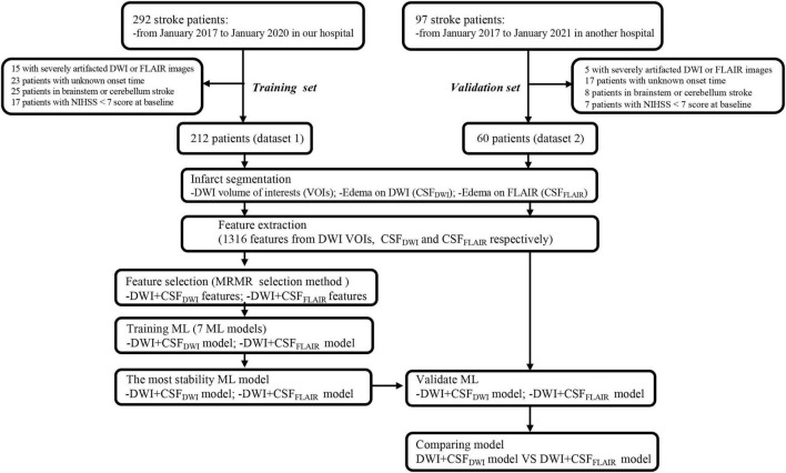
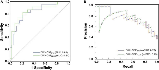
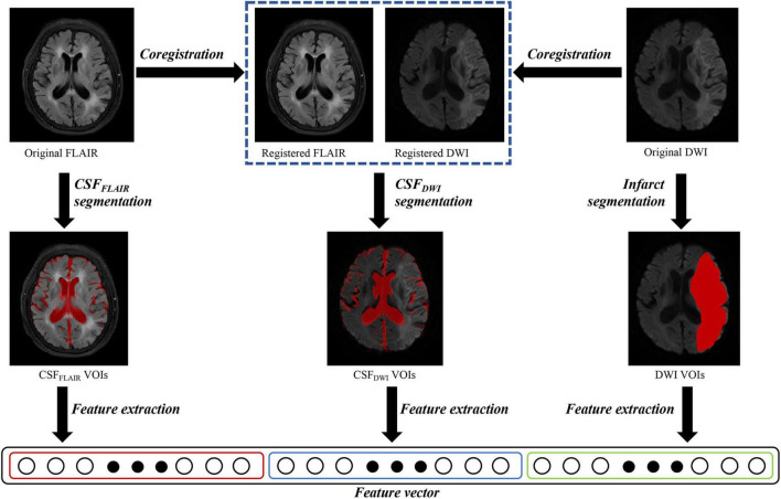
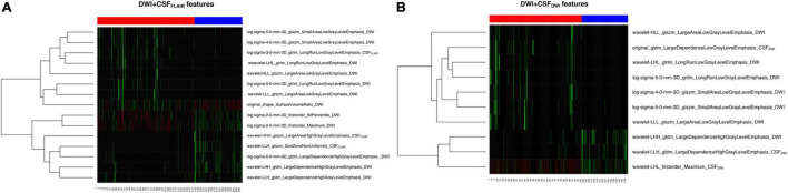
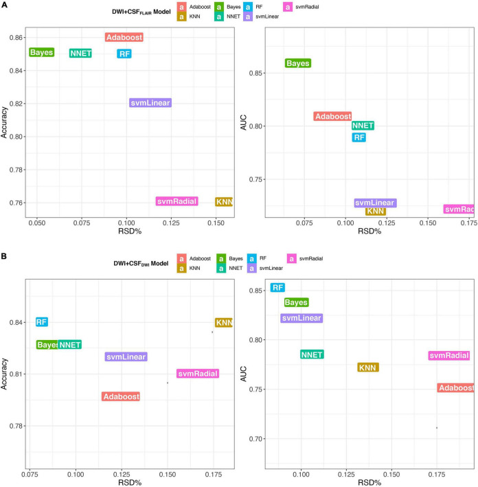
Neuroimaging biomarkers that predict the edema after acute stroke may help clinicians provide targeted therapies and minimize the risk of secondary injury. In this study, we applied pretherapy MRI radiomics features from infarction and cerebrospinal fluid (CSF) to predict edema after acute ischemic stroke. MRI data were obtained from a prospective, endovascular thrombectomy (EVT) cohort that included 389 patients with acute stroke from two centers (dataset 1, = 292; dataset 2, = 97), respectively. Patients were divided into edema group (brain swelling and midline shift) and non-edema group according to CT within 36 h after therapy. We extracted the imaging features of infarct area on diffusion weighted imaging (DWI) (abbreviated as DWI), CSF on fluid-attenuated inversion recovery (FLAIR) (CSF) and CSF on DWI (CSF), and selected the optimum features associated with edema for developing models in two forms of feature sets (DWI + CSF and DWI + CSF) respectively. We developed seven ML models based on dataset 1 and identified the most stable model. External validations (dataset 2) of the developed stable model were performed. Prediction model performance was assessed using the area under the receiver operating characteristic curve (AUC). The Bayes model based on DWI + CSF and the RF model based on DWI + CSF had the best performances (DWI + CSF: AUC, 0.86; accuracy, 0.85; recall, 0.88; DWI + CSF: AUC, 0.86; accuracy, 0.84; recall, 0.84) and the most stability (RSD% in DWI + CSF AUC: 0.07, RSD% in DWI + CSF AUC: 0.09), respectively. External validation showed that the AUC of the Bayes model based on DWI + CSF was 0.84 with accuracy of 0.77 and area under precision-recall curve (auPRC) of 0.75, and the AUC of the RF model based on DWI + CSF was 0.83 with accuracy of 0.81 and the auPRC of 0.76. The MRI radiomics features from infarction and CSF may offer an effective imaging biomarker for predicting edema.

摘要

能够预测急性中风后水肿情况的神经影像学生物标志物,可能有助于临床医生提供针对性治疗,并将继发性损伤风险降至最低。在本研究中,我们应用来自梗死灶和脑脊液(CSF)的治疗前MRI影像组学特征来预测急性缺血性中风后的水肿情况。MRI数据取自一个前瞻性血管内血栓切除术(EVT)队列,该队列分别包含来自两个中心的389例急性中风患者(数据集1,n = 292;数据集2,n = 97)。根据治疗后36小时内的CT检查结果,将患者分为水肿组(脑肿胀和中线移位)和非水肿组。我们提取了扩散加权成像(DWI)上梗死灶区域的影像特征(简称为DWI)、液体衰减反转恢复序列(FLAIR)上的脑脊液(CSF)以及DWI上的脑脊液(CSF),并分别在两种特征集形式(DWI + CSF和DWI + CSF)中选择与水肿相关的最优特征来构建模型。我们基于数据集1开发了七个机器学习模型,并确定了最稳定的模型。对所开发的稳定模型进行了外部验证(数据集2)。使用受试者操作特征曲线下面积(AUC)评估预测模型的性能。基于DWI + CSF的贝叶斯模型和基于DWI + CSF的随机森林(RF)模型分别具有最佳性能(DWI + CSF:AUC为0.86;准确率为0.85;召回率为0.88;DWI + CSF:AUC为0.86;准确率为0.84;召回率为0.84)和最高稳定性(DWI + CSF AUC的相对标准偏差(RSD%):0.07,DWI + CSF AUC的RSD%:0.09)。外部验证表明,基于DWI + CSF的贝叶斯模型的AUC为0.84,准确率为0.77,精确召回率曲线下面积(auPRC)为0.75,基于DWI + CSF的RF模型的AUC为0.83,准确率为0.81,auPRC为0.76。来自梗死灶和脑脊液的MRI影像组学特征可能为预测水肿提供一种有效的影像学生物标志物。

https://cdn.ncbi.nlm.nih.gov/pmc/blobs/a864/8929352/e8577e839bc3/fnagi-14-782036-g005.jpg
https://cdn.ncbi.nlm.nih.gov/pmc/blobs/a864/8929352/52aa007019a1/fnagi-14-782036-g001.jpg
https://cdn.ncbi.nlm.nih.gov/pmc/blobs/a864/8929352/885968e6215c/fnagi-14-782036-g002.jpg
https://cdn.ncbi.nlm.nih.gov/pmc/blobs/a864/8929352/ce95d6ef1c43/fnagi-14-782036-g003.jpg
https://cdn.ncbi.nlm.nih.gov/pmc/blobs/a864/8929352/12c39eacc531/fnagi-14-782036-g004.jpg
https://cdn.ncbi.nlm.nih.gov/pmc/blobs/a864/8929352/e8577e839bc3/fnagi-14-782036-g005.jpg

相似文献

1
MRI Radiomics Features From Infarction and Cerebrospinal Fluid for Prediction of Cerebral Edema After Acute Ischemic Stroke.基于梗死灶和脑脊液的MRI影像组学特征预测急性缺血性卒中后脑水肿
Front Aging Neurosci. 2022 Mar 3;14:782036. doi: 10.3389/fnagi.2022.782036. eCollection 2022.
2
Development and external validation of a stability machine learning model to identify wake-up stroke onset time from MRI.开发并外部验证一种稳定性机器学习模型,以从 MRI 中识别卒中发作的唤醒时间。
Eur Radiol. 2022 Jun;32(6):3661-3669. doi: 10.1007/s00330-021-08493-6. Epub 2022 Jan 17.
3
Prediction of recurrence of ischemic stroke within 1 year of discharge based on machine learning MRI radiomics.基于机器学习MRI影像组学对缺血性卒中出院后1年内复发的预测
Front Neurosci. 2023 May 4;17:1110579. doi: 10.3389/fnins.2023.1110579. eCollection 2023.
4
A deep learning-based model for prediction of hemorrhagic transformation after stroke.基于深度学习的脑卒中后出血性转化预测模型。
Brain Pathol. 2023 Mar;33(2):e13023. doi: 10.1111/bpa.13023. Epub 2021 Oct 4.
5
Quantitative Serial CT Imaging-Derived Features Improve Prediction of Malignant Cerebral Edema after Ischemic Stroke.定量连续 CT 成像衍生特征可提高对缺血性脑卒中后恶性脑水肿的预测能力。
Neurocrit Care. 2020 Dec;33(3):785-792. doi: 10.1007/s12028-020-01056-5. Epub 2020 Jul 29.
6
Prognosis of ischemic stroke predicted by machine learning based on multi-modal MRI radiomics.基于多模态MRI影像组学的机器学习对缺血性中风的预后预测
Front Psychiatry. 2023 Jan 9;13:1105496. doi: 10.3389/fpsyt.2022.1105496. eCollection 2022.
7
Apparent diffusion coefficient map based radiomics model in identifying the ischemic penumbra in acute ischemic stroke.基于表观扩散系数图的放射组学模型在急性缺血性卒中缺血半暗带识别中的应用
Ann Palliat Med. 2020 Sep;9(5):2684-2692. doi: 10.21037/apm-20-1142. Epub 2020 Jul 24.
8
Predicting long-term outcomes for acute ischemic stroke using multi-model MRI radiomics and clinical variables.使用多模态MRI影像组学和临床变量预测急性缺血性卒中的长期预后
Front Med (Lausanne). 2024 Mar 1;11:1328073. doi: 10.3389/fmed.2024.1328073. eCollection 2024.
9
Accelerating Prediction of Malignant Cerebral Edema After Ischemic Stroke with Automated Image Analysis and Explainable Neural Networks.利用自动化图像分析和可解释神经网络加速缺血性脑卒中后恶性脑水肿的预测。
Neurocrit Care. 2022 Apr;36(2):471-482. doi: 10.1007/s12028-021-01325-x. Epub 2021 Aug 20.
10
Combining machine learning with radiomics features in predicting outcomes after mechanical thrombectomy in patients with acute ischemic stroke.将机器学习与放射组学特征相结合,预测急性缺血性脑卒中患者机械取栓治疗后的结局。
Comput Methods Programs Biomed. 2022 Oct;225:107093. doi: 10.1016/j.cmpb.2022.107093. Epub 2022 Aug 28.

引用本文的文献

1
Predicting malignant cerebral edema after acute ischemic stroke: a machine-learning model with multi-region radiomics.预测急性缺血性卒中后恶性脑水肿:一种基于多区域放射组学的机器学习模型
Quant Imaging Med Surg. 2025 Jun 6;15(6):5188-5203. doi: 10.21037/qims-2024-2751. Epub 2025 Jun 3.
2
Predictive Value of Machine Learning Models for Cerebral Edema Risk in Stroke Patients: A Meta-Analysis.机器学习模型对中风患者脑水肿风险的预测价值:一项荟萃分析。
Brain Behav. 2025 Jan;15(1):e70198. doi: 10.1002/brb3.70198.
3
An integrated nomogram combining clinical and radiomic features of hyperattenuated imaging markers to predict malignant cerebral edema following endovascular thrombectomy.

本文引用的文献

1
An automatic machine learning approach for ischemic stroke onset time identification based on DWI and FLAIR imaging.一种基于 DWI 和 FLAIR 成像的缺血性脑卒中发病时间自动识别的机器学习方法。
Neuroimage Clin. 2021;31:102744. doi: 10.1016/j.nicl.2021.102744. Epub 2021 Jul 3.
2
Random Forest-Based Prediction of Outcome and Mortality in Patients with Traumatic Brain Injury Undergoing Primary Decompressive Craniectomy.基于随机森林的原发性去骨瓣减压术后创伤性脑损伤患者预后和死亡率预测。
World Neurosurg. 2021 Apr;148:e450-e458. doi: 10.1016/j.wneu.2021.01.002. Epub 2021 Jan 12.
3
Prediction of Stroke Outcome Using Natural Language Processing-Based Machine Learning of Radiology Report of Brain MRI.
一种结合高衰减成像标志物的临床和影像组学特征的综合列线图,用于预测血管内血栓切除术后的恶性脑水肿。
Quant Imaging Med Surg. 2024 Jul 1;14(7):4936-4949. doi: 10.21037/qims-24-99. Epub 2024 Jun 27.
4
Texture analysis of apparent diffusion coefficient maps in predicting the clinical functional outcomes of acute ischemic stroke.表观扩散系数图的纹理分析在预测急性缺血性卒中临床功能结局中的应用
Front Neurol. 2023 May 11;14:1132318. doi: 10.3389/fneur.2023.1132318. eCollection 2023.
5
Prognostic Value of Combined Radiomic Features from Follow-Up DWI and T2-FLAIR in Acute Ischemic Stroke.随访扩散加权成像(DWI)和液体衰减反转恢复序列(T2-FLAIR)联合影像组学特征在急性缺血性卒中中的预后价值
J Cardiovasc Dev Dis. 2022 Dec 19;9(12):468. doi: 10.3390/jcdd9120468.
6
Diffusion-weighted imaging-based radiomics for predicting 1-year ischemic stroke recurrence.基于扩散加权成像的影像组学用于预测缺血性卒中1年复发情况
Front Neurol. 2022 Oct 28;13:1012896. doi: 10.3389/fneur.2022.1012896. eCollection 2022.
使用基于自然语言处理的脑磁共振成像放射学报告机器学习预测卒中结局
J Pers Med. 2020 Dec 16;10(4):286. doi: 10.3390/jpm10040286.
4
A Nomogram Model to Predict Malignant Cerebral Edema in Ischemic Stroke Patients Treated with Endovascular Thrombectomy: An Observational Study.一种预测接受血管内血栓切除术治疗的缺血性中风患者恶性脑水肿的列线图模型:一项观察性研究。
Neuropsychiatr Dis Treat. 2020 Dec 2;16:2913-2920. doi: 10.2147/NDT.S279303. eCollection 2020.
5
Machine-learning-based outcome prediction in stroke patients with middle cerebral artery-M1 occlusions and early thrombectomy.基于机器学习的大脑中动脉 M1 段闭塞和早期取栓的卒中患者预后预测。
Eur J Neurol. 2021 Apr;28(4):1234-1243. doi: 10.1111/ene.14651. Epub 2020 Dec 21.
6
Machine Learning for Brain Stroke: A Review.机器学习在脑卒中领域的应用:综述。
J Stroke Cerebrovasc Dis. 2020 Oct;29(10):105162. doi: 10.1016/j.jstrokecerebrovasdis.2020.105162. Epub 2020 Jul 28.
7
Interpreting SVM for medical images using Quadtree.使用四叉树对医学图像进行支持向量机解释。
Multimed Tools Appl. 2020;79(39-40):29353-29373. doi: 10.1007/s11042-020-09431-2. Epub 2020 Aug 11.
8
Radiomics in medical imaging-"how-to" guide and critical reflection.医学影像中的放射组学——“操作指南”与批判性思考
Insights Imaging. 2020 Aug 12;11(1):91. doi: 10.1186/s13244-020-00887-2.
9
Machine Learning Prediction of Stroke Mechanism in Embolic Strokes of Undetermined Source.机器学习预测不明来源栓塞性脑卒中的卒中机制。
Stroke. 2020 Sep;51(9):e203-e210. doi: 10.1161/STROKEAHA.120.029305. Epub 2020 Aug 12.
10
Quantitative Serial CT Imaging-Derived Features Improve Prediction of Malignant Cerebral Edema after Ischemic Stroke.定量连续 CT 成像衍生特征可提高对缺血性脑卒中后恶性脑水肿的预测能力。
Neurocrit Care. 2020 Dec;33(3):785-792. doi: 10.1007/s12028-020-01056-5. Epub 2020 Jul 29.