• 文献检索
  • 文档翻译
  • 深度研究
  • 学术资讯
  • Suppr Zotero 插件Zotero 插件
  • 邀请有礼
  • 套餐&价格
  • 历史记录
应用&插件
Suppr Zotero 插件Zotero 插件浏览器插件Mac 客户端Windows 客户端微信小程序
定价
高级版会员购买积分包购买API积分包
服务
文献检索文档翻译深度研究API 文档MCP 服务
关于我们
关于 Suppr公司介绍联系我们用户协议隐私条款
关注我们

Suppr 超能文献

核心技术专利:CN118964589B侵权必究
粤ICP备2023148730 号-1Suppr @ 2026

文献检索

告别复杂PubMed语法,用中文像聊天一样搜索,搜遍4000万医学文献。AI智能推荐,让科研检索更轻松。

立即免费搜索

文件翻译

保留排版,准确专业,支持PDF/Word/PPT等文件格式,支持 12+语言互译。

免费翻译文档

深度研究

AI帮你快速写综述,25分钟生成高质量综述,智能提取关键信息,辅助科研写作。

立即免费体验

不同分割方法对冠状动脉周围脂肪组织诊断性能的影响。

Influence of Different Segmentations on the Diagnostic Performance of Pericoronary Adipose Tissue.

作者信息

Wen Didi, An Rui, Lin Shushen, Yang Wangwei, Jia Yuyang, Zheng Minwen

机构信息

Department of Radiology, Xijing Hospital, Fourth Military Medical University, Xi'an, China.

Siemens Healthineers Ltd., Shanghai, China.

出版信息

Front Cardiovasc Med. 2022 Mar 3;9:773524. doi: 10.3389/fcvm.2022.773524. eCollection 2022.

DOI:10.3389/fcvm.2022.773524
PMID:35310984
原文链接:https://pmc.ncbi.nlm.nih.gov/articles/PMC8929663/
Abstract

OBJECTIVE

To investigate the influence of different segmentations on the diagnostic performance of pericoronary adipose tissue (PCAT) CT attenuation and radiomics features for the prediction of ischemic coronary artery stenosis.

METHODS

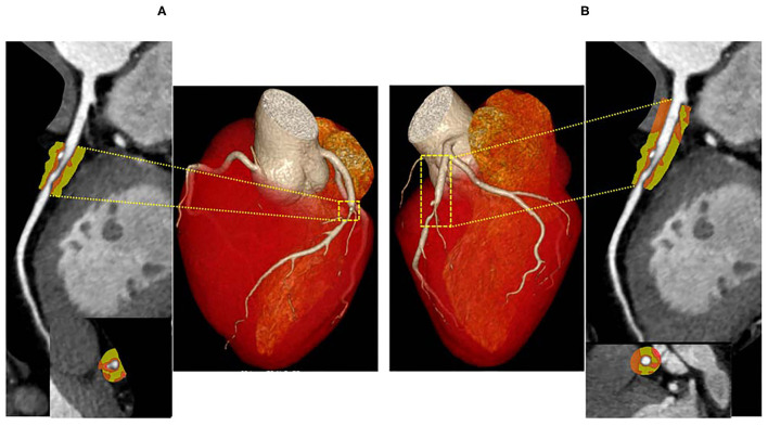
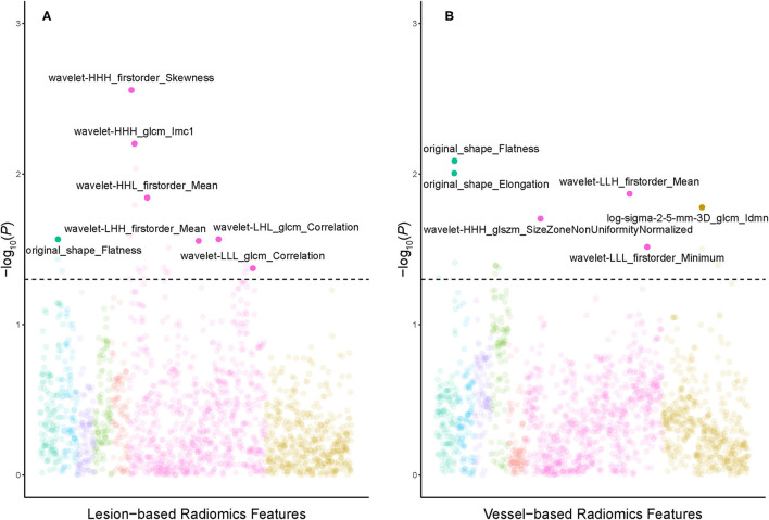
From June 2016 to December 2018, 108 patients with 135 vessels were retrospectively analyzed in the present study. Vessel-based PCAT was segmented along the 40 mm-long proximal segments of three major epicardial coronary arteries, while lesion-based PCAT was defined around coronary lesions. CT attenuation and radiomics features derived from two segmentations were calculated and extracted. The diagnostic performance of PCAT CT attenuation or radiomics models in predicting ischemic coronary stenosis were also compared between vessel-based and lesion-based segmentations.

RESULTS

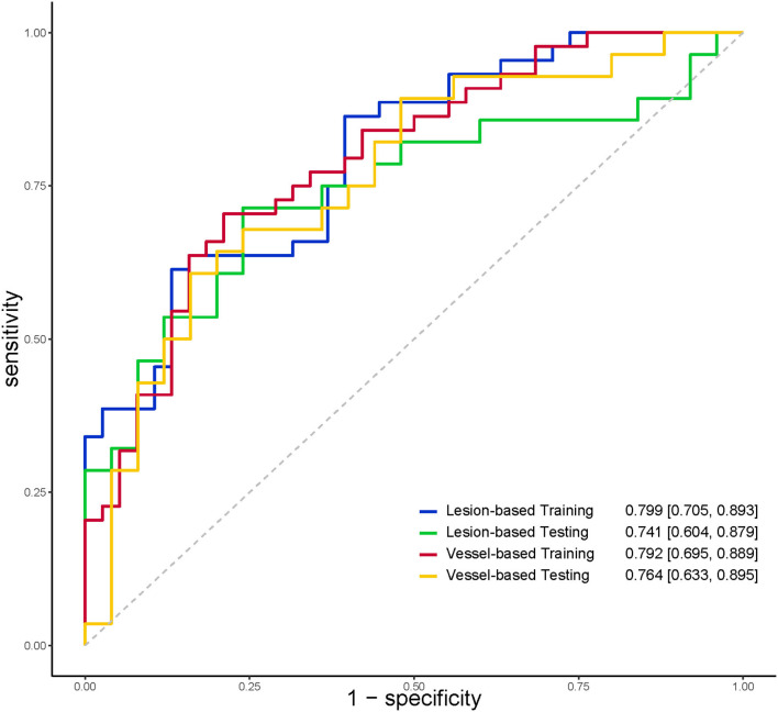
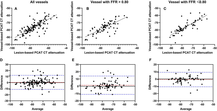
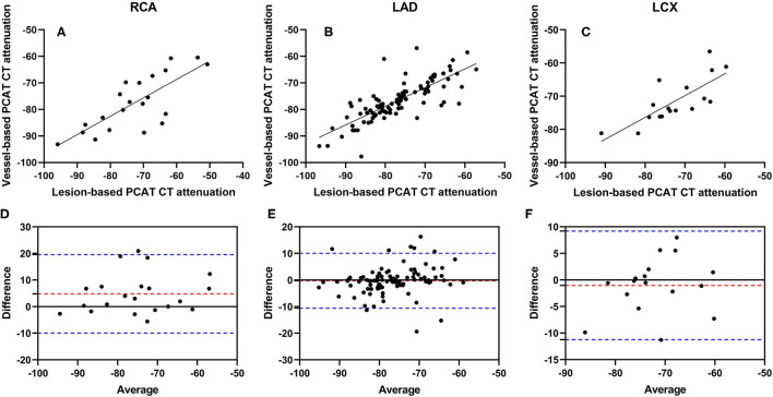
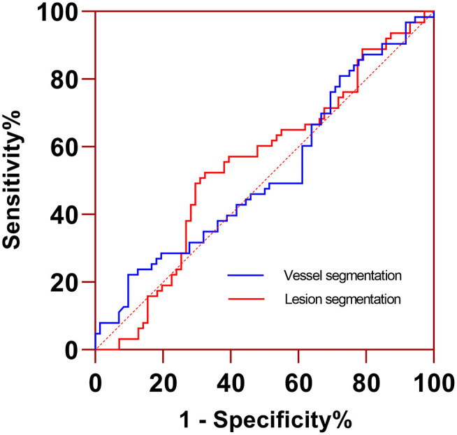
The mean PCAT CT attenuation was -75.7 ± 9.1 HU and -76.1 ± 8.1 HU ( = 0.395) for lesion-based and vessel-based segmentations, respectively. A strong correlation was found between vessel-based and lesion-based PCAT CT attenuation for all cohort and subgroup analyses (all < 0.01). A good agreement for all cohort and subgroup analyses was also detected between two segmentations. The diagnostic performance was comparable between vessel-based and lesion based PCAT CT attenuation in predicting ischemic stenosis. The radiomics features of PCAT based on vessel or lesion segmentation can both adequately identify the ischemic stenosis. However, no significant difference was detected between the two segmentations.

CONCLUSIONS

The quantitative evaluation of PCAT can be reliably measured both from vessel-based and lesion-based segmentation. Furthermore, the radiomics analysis of PCAT may potentially help predict hemodynamically significant coronary artery stenosis.

摘要

目的

探讨不同分割方法对冠状动脉周围脂肪组织(PCAT)CT衰减及影像组学特征在预测缺血性冠状动脉狭窄诊断性能方面的影响。

方法

本研究回顾性分析了2016年6月至2018年12月期间108例患者的135条血管。基于血管的PCAT沿着三条主要心外膜冠状动脉40毫米长的近端节段进行分割,而基于病变的PCAT则定义在冠状动脉病变周围。计算并提取了两种分割方法得出的CT衰减及影像组学特征。还比较了基于血管和基于病变的分割方法在PCAT CT衰减或影像组学模型预测缺血性冠状动脉狭窄方面的诊断性能。

结果

基于病变和基于血管的分割方法的平均PCAT CT衰减分别为-75.7±9.1 HU和-76.1±8.1 HU(P = 0.395)。在所有队列和亚组分析中,基于血管和基于病变的PCAT CT衰减之间均发现有很强的相关性(所有P<0.01)。两种分割方法在所有队列和亚组分析中也显示出良好的一致性。在预测缺血性狭窄方面,基于血管和基于病变的PCAT CT衰减的诊断性能相当。基于血管或病变分割的PCAT影像组学特征均能充分识别缺血性狭窄。然而,两种分割方法之间未检测到显著差异。

结论

PCAT的定量评估可通过基于血管和基于病变的分割方法可靠地进行测量。此外,PCAT的影像组学分析可能有助于预测具有血流动力学意义的冠状动脉狭窄。

https://cdn.ncbi.nlm.nih.gov/pmc/blobs/cef8/8929663/d8a8bcd5aad2/fcvm-09-773524-g0006.jpg
https://cdn.ncbi.nlm.nih.gov/pmc/blobs/cef8/8929663/6d8029228e2f/fcvm-09-773524-g0001.jpg
https://cdn.ncbi.nlm.nih.gov/pmc/blobs/cef8/8929663/b27a2cb10927/fcvm-09-773524-g0002.jpg
https://cdn.ncbi.nlm.nih.gov/pmc/blobs/cef8/8929663/8e004539b28c/fcvm-09-773524-g0003.jpg
https://cdn.ncbi.nlm.nih.gov/pmc/blobs/cef8/8929663/1ba3dbe4db1f/fcvm-09-773524-g0004.jpg
https://cdn.ncbi.nlm.nih.gov/pmc/blobs/cef8/8929663/d2acfb2bc348/fcvm-09-773524-g0005.jpg
https://cdn.ncbi.nlm.nih.gov/pmc/blobs/cef8/8929663/d8a8bcd5aad2/fcvm-09-773524-g0006.jpg

相似文献

1
Influence of Different Segmentations on the Diagnostic Performance of Pericoronary Adipose Tissue.不同分割方法对冠状动脉周围脂肪组织诊断性能的影响。
Front Cardiovasc Med. 2022 Mar 3;9:773524. doi: 10.3389/fcvm.2022.773524. eCollection 2022.
2
Radiomics features of pericoronary adipose tissue improve CT-FFR performance in predicting hemodynamically significant coronary artery stenosis.冠状脂肪组织的放射组学特征可提高 CT-FFR 预测有意义的冠状动脉狭窄的性能。
Eur Radiol. 2023 Mar;33(3):2004-2014. doi: 10.1007/s00330-022-09175-7. Epub 2022 Oct 18.
3
Pericoronary adipose tissue CT attenuation and volume: Diagnostic performance for hemodynamically significant stenosis in patients with suspected coronary artery disease.冠状动脉周围脂肪组织 CT 衰减值和体积:对疑似冠心病患者中具有血流动力学意义的狭窄的诊断性能。
Eur J Radiol. 2021 Jul;140:109740. doi: 10.1016/j.ejrad.2021.109740. Epub 2021 Apr 28.
4
Incremental diagnostic value of radiomics signature of pericoronary adipose tissue for detecting functional myocardial ischemia: a multicenter study.基于冠状动脉脂肪组织影像组学特征对功能性心肌缺血的诊断价值:一项多中心研究。
Eur Radiol. 2023 May;33(5):3007-3019. doi: 10.1007/s00330-022-09377-z. Epub 2023 Feb 2.
5
Non-contrast CT-based radiomics nomogram of pericoronary adipose tissue for predicting haemodynamically significant coronary stenosis in patients with type 2 diabetes.基于非对比 CT 的冠状动脉周围脂肪组织放射组学列线图预测 2 型糖尿病患者的血流动力学意义重大的冠状动脉狭窄。
BMC Med Imaging. 2023 Jul 28;23(1):99. doi: 10.1186/s12880-023-01051-0.
6
Pericoronary Adipose Tissue Computed Tomography Attenuation and High-Risk Plaque Characteristics in Acute Coronary Syndrome Compared With Stable Coronary Artery Disease.对比稳定型冠心病,急性冠状动脉综合征患者的冠状动脉旁脂肪组织 CT 衰减值与高危斑块特征。
JAMA Cardiol. 2018 Sep 1;3(9):858-863. doi: 10.1001/jamacardio.2018.1997.
7
Myocardial Infarction Associates With a Distinct Pericoronary Adipose Tissue Radiomic Phenotype: A Prospective Case-Control Study.心肌梗死与独特的冠状动脉周围脂肪组织放射组学表型相关:一项前瞻性病例对照研究。
JACC Cardiovasc Imaging. 2020 Nov;13(11):2371-2383. doi: 10.1016/j.jcmg.2020.06.033. Epub 2020 Aug 26.
8
Coronary CTA-based radiomic signature of pericoronary adipose tissue predict rapid plaque progression.基于冠状动脉CTA的冠状动脉周围脂肪组织影像组学特征可预测斑块快速进展。
Insights Imaging. 2024 Jun 20;15(1):151. doi: 10.1186/s13244-024-01731-7.
9
Pericoronary fat volume but not attenuation differentiates culprit lesions in patients with myocardial infarction.冠状动脉脂肪体积而非衰减可区分心肌梗死患者的罪犯病变。
Atherosclerosis. 2018 Sep;276:182-188. doi: 10.1016/j.atherosclerosis.2018.05.035. Epub 2018 May 25.
10
Identification of patients with acute myocardial infarction based on coronary CT angiography: the value of pericoronary adipose tissue radiomics.基于冠状动脉 CT 血管造影的急性心肌梗死患者识别:心包脂肪组织放射组学的价值。
Eur Radiol. 2022 Oct;32(10):6868-6877. doi: 10.1007/s00330-022-08812-5. Epub 2022 May 4.

引用本文的文献

1
Predicting chronic stenosis progression of stable coronary artery disease using peri-coronary fat attenuation index derived from coronary computed tomography angiography.利用冠状动脉计算机断层扫描血管造影术得出的冠状动脉周围脂肪衰减指数预测稳定型冠状动脉疾病的慢性狭窄进展
Quant Imaging Med Surg. 2025 Aug 1;15(8):7323-7337. doi: 10.21037/qims-2024-2479. Epub 2025 Jul 25.
2
Cardiac CT Perfusion Imaging of Pericoronary Adipose Tissue (PCAT) Highlighting Potential Confounds in CTA Analysis.冠状动脉周围脂肪组织(PCAT)的心脏CT灌注成像突出了CTA分析中潜在的混杂因素。
J Clin Med. 2025 Jan 24;14(3):769. doi: 10.3390/jcm14030769.
3

本文引用的文献

1
Importance of measurement site on assessment of lesion-specific ischemia and diagnostic performance by coronary computed tomography Angiography-Derived Fractional Flow Reserve.重要性的测量部位评估的特定病变缺血和诊断性能的冠状动脉计算机断层血管造影衍生的分数血流量储备。
J Cardiovasc Comput Tomogr. 2021 Mar-Apr;15(2):114-120. doi: 10.1016/j.jcct.2020.08.005. Epub 2020 Aug 29.
2
Myocardial Infarction Associates With a Distinct Pericoronary Adipose Tissue Radiomic Phenotype: A Prospective Case-Control Study.心肌梗死与独特的冠状动脉周围脂肪组织放射组学表型相关:一项前瞻性病例对照研究。
JACC Cardiovasc Imaging. 2020 Nov;13(11):2371-2383. doi: 10.1016/j.jcmg.2020.06.033. Epub 2020 Aug 26.
3
Pericoronary adipose tissue feature analysis in computed tomography calcium score images in comparison to coronary computed tomography angiography.
计算机断层扫描钙评分图像中冠状动脉周围脂肪组织特征分析与冠状动脉计算机断层扫描血管造影的比较
J Med Imaging (Bellingham). 2025 Jan;12(1):014503. doi: 10.1117/1.JMI.12.1.014503. Epub 2025 Jan 24.
4
Radiomics Signature of Epicardial Adipose Tissue for Predicting Postoperative Atrial Fibrillation after Off-Pump Coronary Artery Bypass Surgery.用于预测非体外循环冠状动脉搭桥术后房颤的心外膜脂肪组织的影像组学特征
Rev Cardiovasc Med. 2023 Nov 23;24(11):327. doi: 10.31083/j.rcm2411327. eCollection 2023 Nov.
5
Radiomics analysis of lesion-specific pericoronary adipose tissue to predict major adverse cardiovascular events in coronary artery disease.基于病变特异性冠状动脉周围脂肪组织的影像组学分析预测冠心病患者的主要不良心血管事件。
BMC Med Imaging. 2024 Jun 17;24(1):150. doi: 10.1186/s12880-024-01325-1.
6
Epicardial adipose tissue, metabolic disorders, and cardiovascular diseases: recent advances classified by research methodologies.心外膜脂肪组织、代谢紊乱与心血管疾病:基于研究方法的最新进展
MedComm (2020). 2023 Oct 24;4(6):e413. doi: 10.1002/mco2.413. eCollection 2023 Dec.
Feasibility of measuring pericoronary fat from precontrast scans: Effect of iodinated contrast on pericoronary fat attenuation.
从平扫图像测量冠状动脉脂肪的可行性:碘对比剂对冠状动脉脂肪衰减的影响。
J Cardiovasc Comput Tomogr. 2020 Nov-Dec;14(6):490-494. doi: 10.1016/j.jcct.2020.04.004. Epub 2020 Apr 22.
4
Repeatability of quantitative pericoronary adipose tissue attenuation and coronary plaque burden from coronary CT angiography.冠状动脉 CT 血管造影定量评估冠状动脉周围脂肪组织衰减和冠状动脉斑块负担的可重复性。
J Cardiovasc Comput Tomogr. 2021 Jan-Feb;15(1):81-84. doi: 10.1016/j.jcct.2020.03.007. Epub 2020 Apr 14.
5
Association between perivascular inflammation and downstream myocardial perfusion in patients with suspected coronary artery disease.血管周围炎症与疑似冠心病患者下游心肌灌注的关系。
Eur Heart J Cardiovasc Imaging. 2020 Jun 1;21(6):599-605. doi: 10.1093/ehjci/jeaa023.
6
Pericoronary adipose tissue and quantitative global non-calcified plaque characteristics from CT angiography do not differ in matched South Asian, East Asian and European-origin Caucasian patients with stable chest pain.经 CT 血管造影检查,在有稳定胸痛的匹配南亚裔、东亚裔和欧洲裔白种人患者中,冠状动脉周围脂肪组织和定量整体非钙化斑块特征并无差异。
Eur J Radiol. 2020 Apr;125:108874. doi: 10.1016/j.ejrad.2020.108874. Epub 2020 Feb 7.
7
Deep Learning-Based Quantification of Epicardial Adipose Tissue Volume and Attenuation Predicts Major Adverse Cardiovascular Events in Asymptomatic Subjects.基于深度学习的心外膜脂肪组织体积和衰减定量预测无症状患者的主要不良心血管事件。
Circ Cardiovasc Imaging. 2020 Feb;13(2):e009829. doi: 10.1161/CIRCIMAGING.119.009829. Epub 2020 Feb 17.
8
Peri-coronary inflammation is associated with findings on coronary computed tomography angiography and fractional flow reserve.冠状动脉周围炎症与冠状动脉计算机断层血管造影和血流储备分数的发现有关。
J Cardiovasc Comput Tomogr. 2020 Nov-Dec;14(6):483-489. doi: 10.1016/j.jcct.2020.02.002. Epub 2020 Feb 6.
9
Clinical Outcomes of Dynamic Computed Tomography Myocardial Perfusion Imaging Combined With Coronary Computed Tomography Angiography Versus Coronary Computed Tomography Angiography-Guided Strategy.动态计算机断层扫描心肌灌注成像联合冠状动脉计算机断层血管造影与冠状动脉计算机断层血管造影指导策略的临床结果。
Circ Cardiovasc Imaging. 2020 Jan;13(1):e009775. doi: 10.1161/CIRCIMAGING.119.009775. Epub 2020 Jan 8.
10
Perivascular fat attenuation index and high-risk plaque features evaluated by coronary CT angiography: relationship with serum inflammatory marker level.冠状动脉 CT 血管造影评估血管周围脂肪衰减指数和高危斑块特征与血清炎症标志物水平的关系。
Int J Cardiovasc Imaging. 2020 Apr;36(4):723-730. doi: 10.1007/s10554-019-01758-8. Epub 2020 Jan 6.