• 文献检索
  • 文档翻译
  • 深度研究
  • 学术资讯
  • Suppr Zotero 插件Zotero 插件
  • 邀请有礼
  • 套餐&价格
  • 历史记录
应用&插件
Suppr Zotero 插件Zotero 插件浏览器插件Mac 客户端Windows 客户端微信小程序
定价
高级版会员购买积分包购买API积分包
服务
文献检索文档翻译深度研究API 文档MCP 服务
关于我们
关于 Suppr公司介绍联系我们用户协议隐私条款
关注我们

Suppr 超能文献

核心技术专利:CN118964589B侵权必究
粤ICP备2023148730 号-1Suppr @ 2026

文献检索

告别复杂PubMed语法,用中文像聊天一样搜索,搜遍4000万医学文献。AI智能推荐,让科研检索更轻松。

立即免费搜索

文件翻译

保留排版,准确专业,支持PDF/Word/PPT等文件格式,支持 12+语言互译。

免费翻译文档

深度研究

AI帮你快速写综述,25分钟生成高质量综述,智能提取关键信息,辅助科研写作。

立即免费体验

配体结合重塑蛋白质侧链构象异质性。

Ligand binding remodels protein side-chain conformational heterogeneity.

机构信息

Department of Bioengineering and Therapeutic Sciences, University of California, San Francisco, San Francisco, United States.

Biophysics Graduate Program, University of California San Francisco, San Francisco, United States.

出版信息

Elife. 2022 Mar 21;11:e74114. doi: 10.7554/eLife.74114.

DOI:10.7554/eLife.74114
PMID:35312477
原文链接:https://pmc.ncbi.nlm.nih.gov/articles/PMC9084896/
Abstract

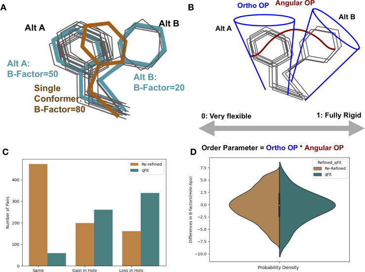
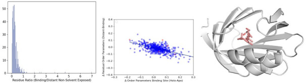
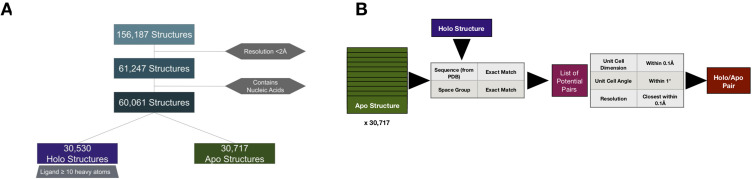
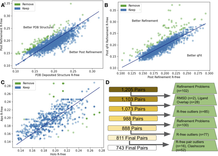
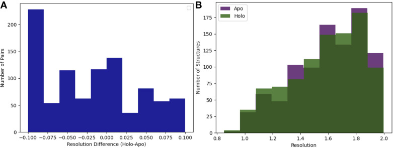
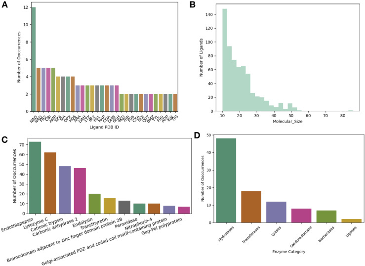
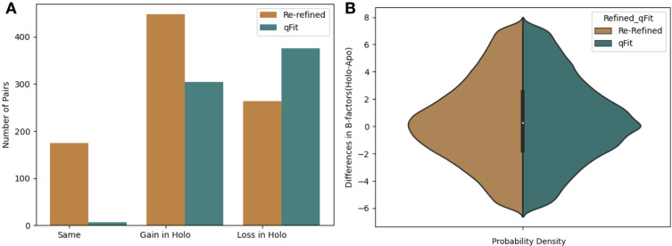
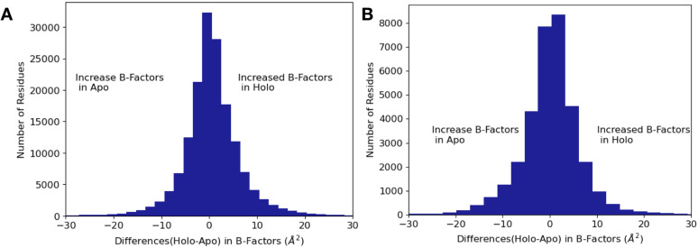
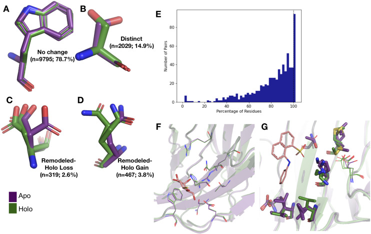
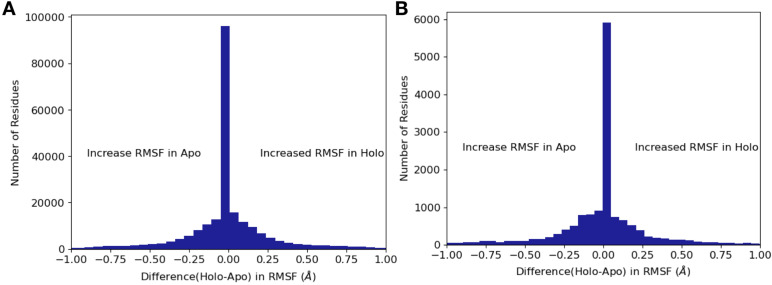
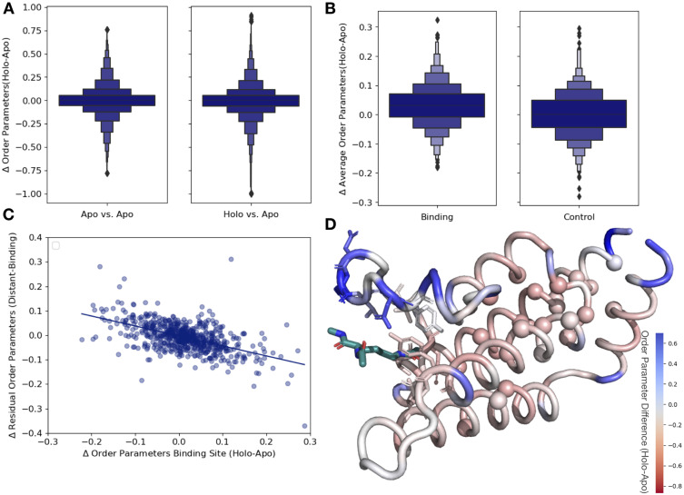
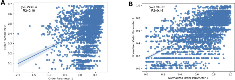
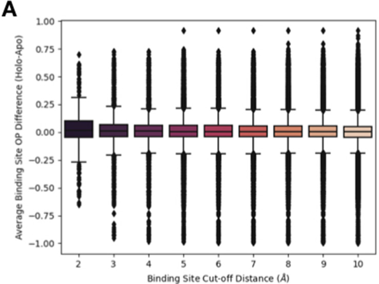
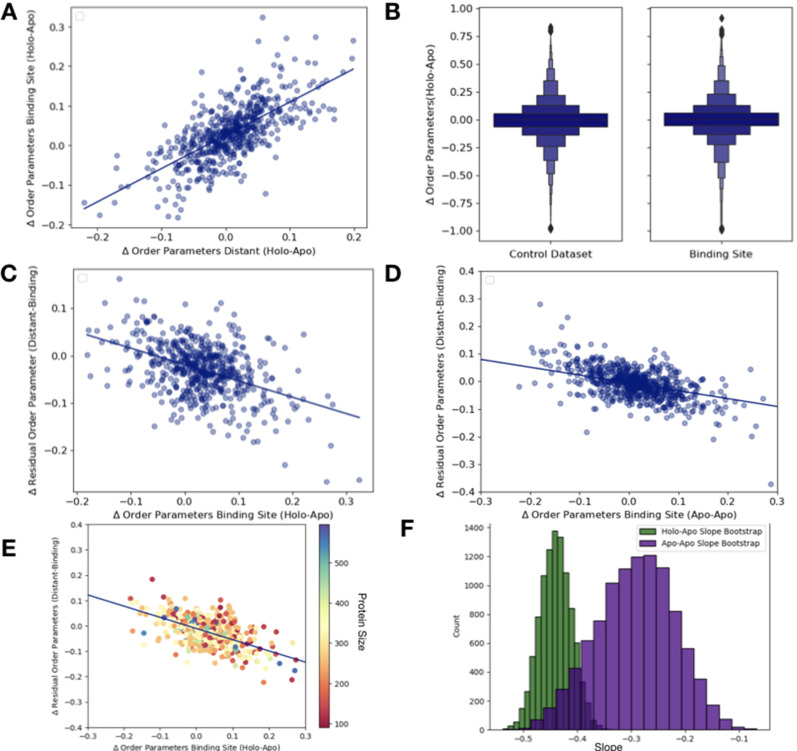
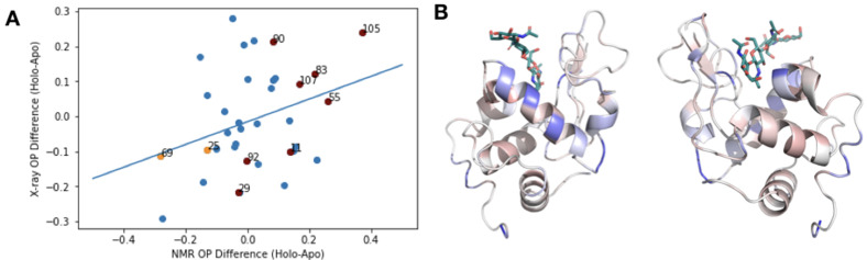
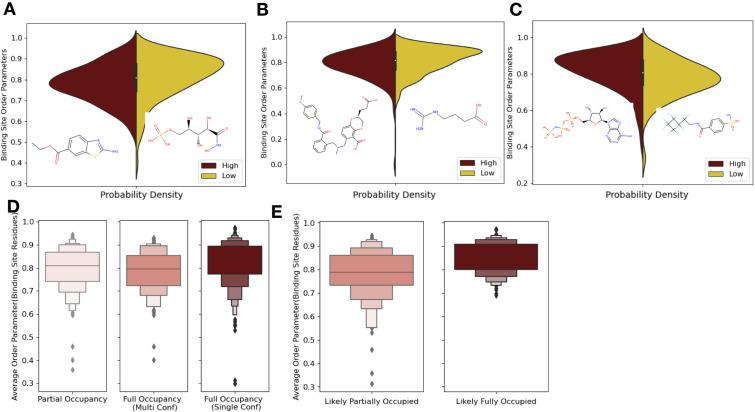
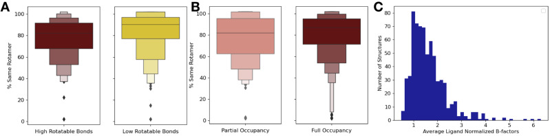
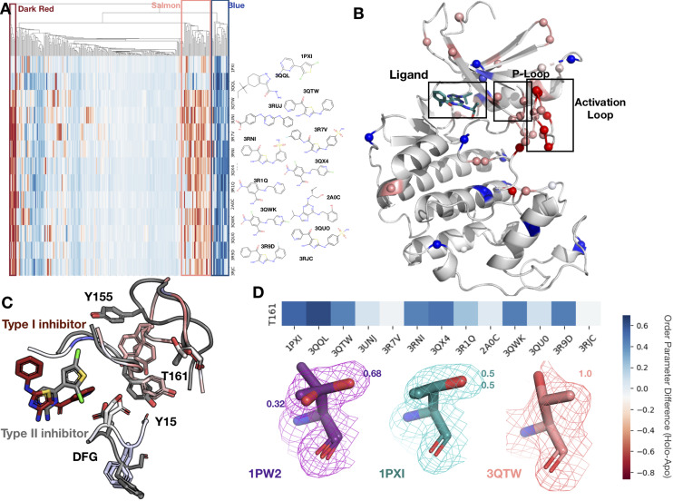
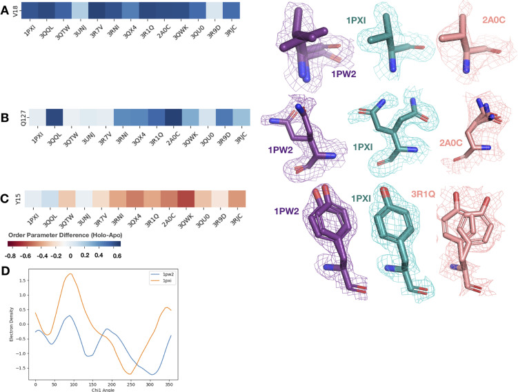
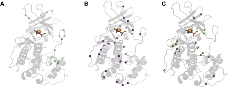
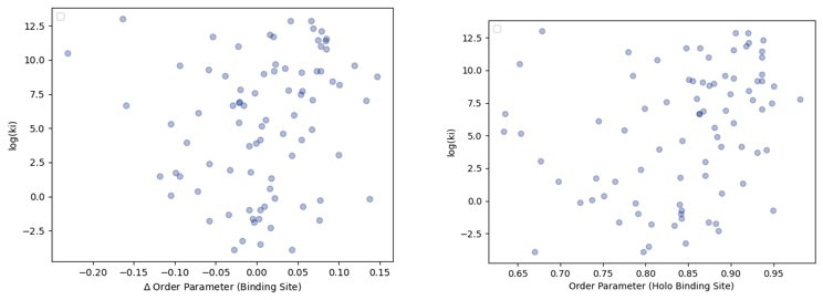
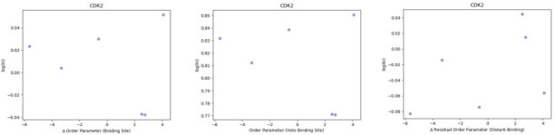
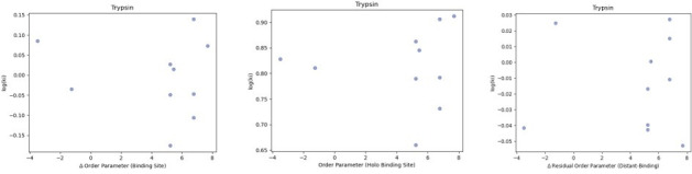
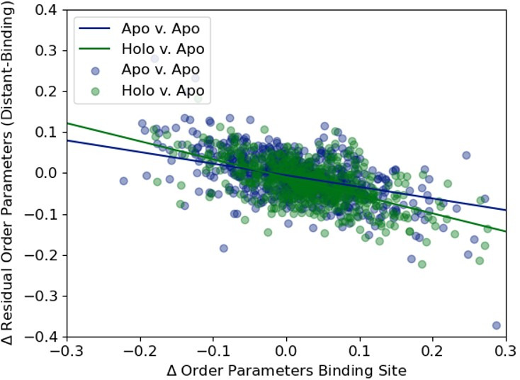
While protein conformational heterogeneity plays an important role in many aspects of biological function, including ligand binding, its impact has been difficult to quantify. Macromolecular X-ray diffraction is commonly interpreted with a static structure, but it can provide information on both the anharmonic and harmonic contributions to conformational heterogeneity. Here, through multiconformer modeling of time- and space-averaged electron density, we measure conformational heterogeneity of 743 stringently matched pairs of crystallographic datasets that reflect unbound/apo and ligand-bound/holo states. When comparing the conformational heterogeneity of side chains, we observe that when binding site residues become more rigid upon ligand binding, distant residues tend to become more flexible, especially in non-solvent-exposed regions. Among ligand properties, we observe increased protein flexibility as the number of hydrogen bonds decreases and relative hydrophobicity increases. Across a series of 13 inhibitor-bound structures of CDK2, we find that conformational heterogeneity is correlated with inhibitor features and identify how conformational changes propagate differences in conformational heterogeneity away from the binding site. Collectively, our findings agree with models emerging from nuclear magnetic resonance studies suggesting that residual side-chain entropy can modulate affinity and point to the need to integrate both static conformational changes and conformational heterogeneity in models of ligand binding.

摘要

虽然蛋白质构象异质性在许多方面都发挥着重要作用,包括配体结合,但它的影响一直难以量化。大分子 X 射线衍射通常采用静态结构进行解释,但它可以提供关于构象异质性的非谐和及调和贡献的信息。在这里,我们通过对时间和空间平均电子密度的多构象建模,测量了 743 对严格匹配的晶体数据集的构象异质性,这些数据集反映了无配体/apo 和配体结合/holo 状态。在比较侧链构象异质性时,我们观察到当结合位点残基在配体结合时变得更加刚性时,远距离残基往往变得更加灵活,特别是在非溶剂暴露区域。在一系列 13 个 CDK2 抑制剂结合结构中,我们发现构象异质性与抑制剂特征相关,并确定构象变化如何将构象异质性的差异从结合位点传播开来。总的来说,我们的发现与核磁共振研究中出现的模型一致,表明残侧链熵可以调节亲和力,并指出需要在配体结合模型中整合静态构象变化和构象异质性。

https://cdn.ncbi.nlm.nih.gov/pmc/blobs/faa1/9084896/83d22eaef290/elife-74114-sa2-fig5.jpg
https://cdn.ncbi.nlm.nih.gov/pmc/blobs/faa1/9084896/71f53d54e780/elife-74114-fig1.jpg
https://cdn.ncbi.nlm.nih.gov/pmc/blobs/faa1/9084896/74bf3ea31409/elife-74114-fig1-figsupp1.jpg
https://cdn.ncbi.nlm.nih.gov/pmc/blobs/faa1/9084896/e88dfd141bb4/elife-74114-fig1-figsupp2.jpg
https://cdn.ncbi.nlm.nih.gov/pmc/blobs/faa1/9084896/1f0092b0e85e/elife-74114-fig1-figsupp3.jpg
https://cdn.ncbi.nlm.nih.gov/pmc/blobs/faa1/9084896/e7a0da20b85a/elife-74114-fig1-figsupp4.jpg
https://cdn.ncbi.nlm.nih.gov/pmc/blobs/faa1/9084896/b2e1348272b8/elife-74114-fig1-figsupp5.jpg
https://cdn.ncbi.nlm.nih.gov/pmc/blobs/faa1/9084896/6bfe128776fb/elife-74114-fig1-figsupp6.jpg
https://cdn.ncbi.nlm.nih.gov/pmc/blobs/faa1/9084896/c4a180f98321/elife-74114-fig2.jpg
https://cdn.ncbi.nlm.nih.gov/pmc/blobs/faa1/9084896/fd7d40151532/elife-74114-fig2-figsupp1.jpg
https://cdn.ncbi.nlm.nih.gov/pmc/blobs/faa1/9084896/f620cb1c05ad/elife-74114-fig3.jpg
https://cdn.ncbi.nlm.nih.gov/pmc/blobs/faa1/9084896/4a9f14519920/elife-74114-fig3-figsupp1.jpg
https://cdn.ncbi.nlm.nih.gov/pmc/blobs/faa1/9084896/4d62d29e9b7b/elife-74114-fig3-figsupp2.jpg
https://cdn.ncbi.nlm.nih.gov/pmc/blobs/faa1/9084896/e5ed157f1b8d/elife-74114-fig3-figsupp3.jpg
https://cdn.ncbi.nlm.nih.gov/pmc/blobs/faa1/9084896/fd5dc6d4c932/elife-74114-fig3-figsupp4.jpg
https://cdn.ncbi.nlm.nih.gov/pmc/blobs/faa1/9084896/d39c62788ad7/elife-74114-fig3-figsupp5.jpg
https://cdn.ncbi.nlm.nih.gov/pmc/blobs/faa1/9084896/168cd125ce7b/elife-74114-fig4.jpg
https://cdn.ncbi.nlm.nih.gov/pmc/blobs/faa1/9084896/0f7e22326434/elife-74114-fig4-figsupp1.jpg
https://cdn.ncbi.nlm.nih.gov/pmc/blobs/faa1/9084896/763fb478cf95/elife-74114-fig5.jpg
https://cdn.ncbi.nlm.nih.gov/pmc/blobs/faa1/9084896/27da2ec4db93/elife-74114-fig5-figsupp1.jpg
https://cdn.ncbi.nlm.nih.gov/pmc/blobs/faa1/9084896/cfea81e08747/elife-74114-fig5-figsupp2.jpg
https://cdn.ncbi.nlm.nih.gov/pmc/blobs/faa1/9084896/2d74d9fa222b/elife-74114-sa2-fig1.jpg
https://cdn.ncbi.nlm.nih.gov/pmc/blobs/faa1/9084896/aeab82dda3b4/elife-74114-sa2-fig2.jpg
https://cdn.ncbi.nlm.nih.gov/pmc/blobs/faa1/9084896/772ef979d7e8/elife-74114-sa2-fig3.jpg
https://cdn.ncbi.nlm.nih.gov/pmc/blobs/faa1/9084896/b8f0a1f20c2c/elife-74114-sa2-fig4.jpg
https://cdn.ncbi.nlm.nih.gov/pmc/blobs/faa1/9084896/83d22eaef290/elife-74114-sa2-fig5.jpg

相似文献

1
Ligand binding remodels protein side-chain conformational heterogeneity.配体结合重塑蛋白质侧链构象异质性。
Elife. 2022 Mar 21;11:e74114. doi: 10.7554/eLife.74114.
2
How different are structurally flexible and rigid binding sites? Sequence and structural features discriminating proteins that do and do not undergo conformational change upon ligand binding.结构灵活和刚性的结合位点有何不同?区分在配体结合时发生和不发生构象变化的蛋白质的序列和结构特征。
J Mol Biol. 2007 Jan 5;365(1):257-73. doi: 10.1016/j.jmb.2006.09.062. Epub 2006 Sep 29.
3
Side-chain rotamer changes upon ligand binding: common, crucial, correlate with entropy and rearrange hydrogen bonding.配体结合导致侧链构象变化:常见、关键、与熵相关、重排氢键。
Bioinformatics. 2012 Sep 15;28(18):i423-i430. doi: 10.1093/bioinformatics/bts395.
4
Macromolecular crowding: chemistry and physics meet biology (Ascona, Switzerland, 10-14 June 2012).大分子拥挤现象:化学与物理邂逅生物学(瑞士阿斯科纳,2012年6月10日至14日)
Phys Biol. 2013 Aug;10(4):040301. doi: 10.1088/1478-3975/10/4/040301. Epub 2013 Aug 2.
5
Inherent versus induced protein flexibility: Comparisons within and between apo and holo structures.固有蛋白与诱导蛋白的柔性:去折叠态和天然态结构内及结构间的比较。
PLoS Comput Biol. 2019 Jan 30;15(1):e1006705. doi: 10.1371/journal.pcbi.1006705. eCollection 2019 Jan.
6
Evidence of conformational selection driving the formation of ligand binding sites in protein-protein interfaces.构象选择驱动蛋白质-蛋白质界面中配体结合位点形成的证据。
PLoS Comput Biol. 2014 Oct 2;10(10):e1003872. doi: 10.1371/journal.pcbi.1003872. eCollection 2014 Oct.
7
Flexible CDOCKER: Hybrid Searching Algorithm and Scoring Function with Side Chain Conformational Entropy.柔性 CDOCKER:带有侧链构象熵的混合搜索算法和打分函数。
J Chem Inf Model. 2021 Nov 22;61(11):5535-5549. doi: 10.1021/acs.jcim.1c01078. Epub 2021 Oct 27.
8
AutoDockFR: Advances in Protein-Ligand Docking with Explicitly Specified Binding Site Flexibility.AutoDockFR:具有明确指定结合位点灵活性的蛋白质-配体对接技术进展
PLoS Comput Biol. 2015 Dec 2;11(12):e1004586. doi: 10.1371/journal.pcbi.1004586. eCollection 2015 Dec.
9
Are induced fit protein conformational changes caused by ligand-binding predictable? A molecular dynamics investigation.配体结合引起的诱导契合蛋白构象变化是否可预测?分子动力学研究。
J Comput Chem. 2017 Jun 5;38(15):1229-1237. doi: 10.1002/jcc.24714. Epub 2017 Apr 16.
10
Side-chain flexibility in proteins upon ligand binding.配体结合后蛋白质侧链的灵活性。
Proteins. 2000 May 15;39(3):261-8. doi: 10.1002/(sici)1097-0134(20000515)39:3<261::aid-prot90>3.0.co;2-4.

引用本文的文献

1
Expanding automated multiconformer ligand modeling to macrocycles and fragments.将自动化多构象配体建模扩展至大环化合物和片段。
Elife. 2025 Jun 30;14:RP103797. doi: 10.7554/eLife.103797.
2
An experimental proxy of water displaceability for ligand discovery.用于配体发现的水置换性实验替代指标。
Nat Methods. 2025 Jun 27. doi: 10.1038/s41592-025-02724-0.
3
Mapping allosteric rewiring in related protein structures from collections of crystallographic multiconformer models.从晶体学多构象体模型集合中映射相关蛋白质结构中的变构重排。

本文引用的文献

1
Exploring ligand dynamics in protein crystal structures with ensemble refinement.利用整体 refinement 方法探索蛋白质晶体结构中的配体动态。
Acta Crystallogr D Struct Biol. 2021 Aug 1;77(Pt 8):1099-1115. doi: 10.1107/S2059798321006513. Epub 2021 Jul 29.
2
The evolution of cyclin dependent kinase inhibitors in the treatment of cancer.细胞周期蛋白依赖性激酶抑制剂在癌症治疗中的演变。
Expert Rev Anticancer Ther. 2021 Oct;21(10):1105-1124. doi: 10.1080/14737140.2021.1944109. Epub 2021 Sep 1.
3
qFit 3: Protein and ligand multiconformer modeling for X-ray crystallographic and single-particle cryo-EM density maps.
bioRxiv. 2025 May 27:2025.05.23.655529. doi: 10.1101/2025.05.23.655529.
4
TRPM8 protein dynamics correlates with ligand structure and cellular function.瞬时受体电位阳离子通道亚家族M成员8(TRPM8)蛋白动力学与配体结构和细胞功能相关。
bioRxiv. 2025 May 15:2025.05.13.653789. doi: 10.1101/2025.05.13.653789.
5
TRPM8 Protein Dynamics Correlates with Ligand Structure and Cellular Function.瞬时受体电位阳离子通道亚家族M成员8(TRPM8)蛋白动力学与配体结构及细胞功能相关。
J Am Chem Soc. 2025 Jun 4;147(22):18460-18474. doi: 10.1021/jacs.4c09435. Epub 2025 May 27.
6
Advances in uncovering the mechanisms of macromolecular conformational entropy.揭示大分子构象熵机制的进展。
Nat Chem Biol. 2025 May;21(5):623-634. doi: 10.1038/s41589-025-01879-3. Epub 2025 Apr 24.
7
AlphaFold as a Prior: Experimental Structure Determination Conditioned on a Pretrained Neural Network.以AlphaFold为先验:基于预训练神经网络的实验结构测定
bioRxiv. 2025 Mar 11:2025.02.18.638828. doi: 10.1101/2025.02.18.638828.
8
FLEXR-MSA: electron-density map comparisons of sequence-diverse structures.FLEXR-MSA:序列多样结构的电子密度图比较
IUCrJ. 2025 Mar 1;12(Pt 2):245-254. doi: 10.1107/S2052252525001332.
9
Proteins with alternative folds reveal blind spots in AlphaFold-based protein structure prediction.具有可变折叠的蛋白质揭示了基于AlphaFold的蛋白质结构预测中的盲点。
ArXiv. 2024 Oct 18:arXiv:2410.14898v1.
10
Proteins with alternative folds reveal blind spots in AlphaFold-based protein structure prediction.具有可变折叠的蛋白质揭示了基于AlphaFold的蛋白质结构预测中的盲点。
Curr Opin Struct Biol. 2025 Feb;90:102973. doi: 10.1016/j.sbi.2024.102973. Epub 2025 Jan 4.
qFit 3:用于 X 射线晶体学和单颗粒冷冻电镜密度图的蛋白质和配体多构象建模。
Protein Sci. 2021 Jan;30(1):270-285. doi: 10.1002/pro.4001. Epub 2020 Nov 30.
4
XGen: Real-Space Fitting of Complex Ligand Conformational Ensembles to X-ray Electron Density Maps.XGen:X 射线电子密度图中复杂配体构象集合的实空间拟合。
J Med Chem. 2020 Sep 24;63(18):10509-10528. doi: 10.1021/acs.jmedchem.0c01373. Epub 2020 Sep 14.
5
Macromolecular structure determination using X-rays, neutrons and electrons: recent developments in Phenix.利用 X 射线、中子和电子进行高分子结构测定: Phenix 的最新进展。
Acta Crystallogr D Struct Biol. 2019 Oct 1;75(Pt 10):861-877. doi: 10.1107/S2059798319011471. Epub 2019 Oct 2.
6
Effects of α-tubulin acetylation on microtubule structure and stability.α-微管蛋白乙酰化对微管结构和稳定性的影响。
Proc Natl Acad Sci U S A. 2019 May 21;116(21):10366-10371. doi: 10.1073/pnas.1900441116. Epub 2019 May 9.
7
Globally correlated conformational entropy underlies positive and negative cooperativity in a kinase's enzymatic cycle.全球相关构象熵是激酶酶循环中正负协同作用的基础。
Nat Commun. 2019 Feb 18;10(1):799. doi: 10.1038/s41467-019-08655-7.
8
Interplay between Conformational Entropy and Solvation Entropy in Protein-Ligand Binding.构象熵和溶剂化熵在蛋白-配体结合中的相互作用。
J Am Chem Soc. 2019 Feb 6;141(5):2012-2026. doi: 10.1021/jacs.8b11099. Epub 2019 Jan 23.
9
qFit-ligand Reveals Widespread Conformational Heterogeneity of Drug-Like Molecules in X-Ray Electron Density Maps.qFit-配体揭示了在 X 射线电子密度图中药物样分子的广泛构象异质性。
J Med Chem. 2018 Dec 27;61(24):11183-11198. doi: 10.1021/acs.jmedchem.8b01292. Epub 2018 Dec 6.
10
Structural insights into the functional diversity of the CDK-cyclin family.结构洞察细胞周期蛋白依赖性激酶-细胞周期蛋白家族的功能多样性。
Open Biol. 2018 Sep;8(9). doi: 10.1098/rsob.180112.