Ogawa Kaho, Ishida Yoshiki, Kuwajima Yukinori, Lee Cliff, Emge Jacob R, Izumisawa Mitsuru, Satoh Kazuro, Ishikawa-Nagai Shigemi, Da Silva John D, Chen Chia-Yu
Department of Oral Medicine, Immunity and Infection, Harvard School of Dental Medicine, 188 Longwood Avenue, Boston, MA 02115, USA.
Division of Orthodontics, Department of Developmental Oral Health Science, School of Dentistry Iwate Medical University, 1-3-27 Chuo-dori, Morioka 020-8505, Iwate, Japan.
Tomography. 2022 Feb 23;8(2):550-559. doi: 10.3390/tomography8020045.
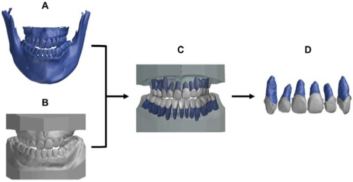
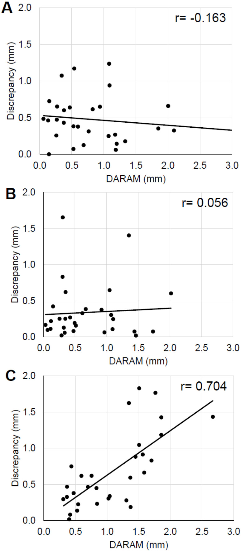
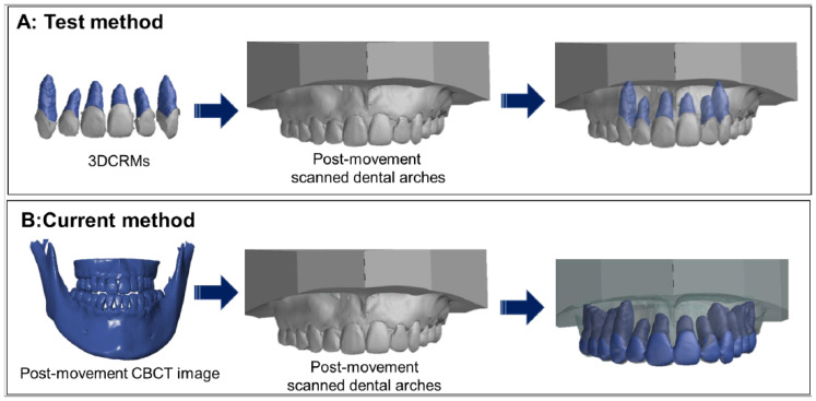
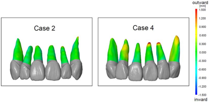
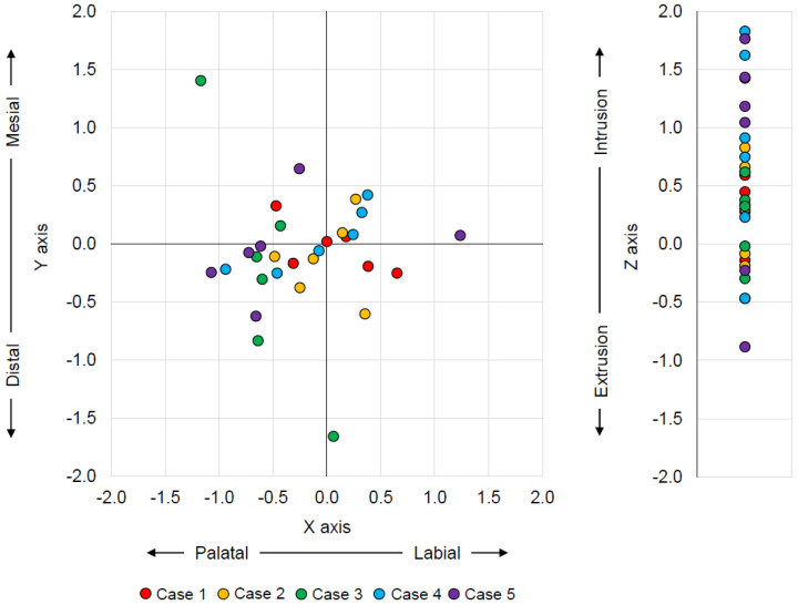
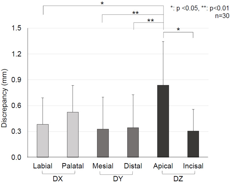
This study aimed to assess the accuracy of a method of predicting post-movement root position during orthodontic treatment using a 3D digital crown/root model (3DCRM) created with pre-movement records of both cone-beam computed tomography (CBCT) and dental arch digital scans. Pre- and post-movement CBCT scans and dental arch digital scans of five patients who had completed orthodontic treatments were used in this study. The 3DCRM was superimposed onto the post-movement scanned dental arch to identify the post-movement root position (test method). Post-movement CBCT (referenced as the current method) served as the control to identify the actual post-movement root position. 3D-coordinate analysis revealed no significant differences between the test and current methods along the X and Y axes. However, the discrepancy on the Z axis (especially in cases of intrusion) was greater than that in all other directions for all three tooth types examined (p < 0.05). A strong positive correlation between the degree of discrepancy and the distance of tooth movement was observed on the Z axis (r = 0.71). The 3DCRM method showed promising potential to accurately predict root position during orthodontic treatments without the need for a second CBCT. However, root resorption, which affected the Z axis prediction, needs to be closely monitored using periapical radiographs to complement this method.
本研究旨在评估一种利用锥形束计算机断层扫描(CBCT)和牙弓数字扫描的移动前记录创建的三维数字冠/根模型(3DCRM)预测正畸治疗中移动后牙根位置方法的准确性。本研究使用了五名完成正畸治疗患者的移动前和移动后CBCT扫描以及牙弓数字扫描。将3DCRM叠加到移动后扫描的牙弓上,以确定移动后牙根位置(测试方法)。移动后CBCT(作为当前方法)作为对照,以确定实际的移动后牙根位置。三维坐标分析显示,测试方法和当前方法在X轴和Y轴上无显著差异。然而,在所检查的所有三种牙齿类型中,Z轴上的差异(尤其是在牙齿压低的情况下)大于所有其他方向(p<0.05)。在Z轴上观察到差异程度与牙齿移动距离之间存在强正相关(r=0.71)。3DCRM方法显示出在正畸治疗期间无需第二次CBCT即可准确预测牙根位置的潜在前景。然而,影响Z轴预测的牙根吸收需要使用根尖片进行密切监测,以补充该方法。