• 文献检索
  • 文档翻译
  • 深度研究
  • 学术资讯
  • Suppr Zotero 插件Zotero 插件
  • 邀请有礼
  • 套餐&价格
  • 历史记录
应用&插件
Suppr Zotero 插件Zotero 插件浏览器插件Mac 客户端Windows 客户端微信小程序
定价
高级版会员购买积分包购买API积分包
服务
文献检索文档翻译深度研究API 文档MCP 服务
关于我们
关于 Suppr公司介绍联系我们用户协议隐私条款
关注我们

Suppr 超能文献

核心技术专利:CN118964589B侵权必究
粤ICP备2023148730 号-1Suppr @ 2026

文献检索

告别复杂PubMed语法,用中文像聊天一样搜索,搜遍4000万医学文献。AI智能推荐,让科研检索更轻松。

立即免费搜索

文件翻译

保留排版,准确专业,支持PDF/Word/PPT等文件格式,支持 12+语言互译。

免费翻译文档

深度研究

AI帮你快速写综述,25分钟生成高质量综述,智能提取关键信息,辅助科研写作。

立即免费体验

天然细胞外囊泡在人诱导多能干细胞向心肌细胞分化中的生物活性和 miRNA 谱分析。

Bioactivity and miRNome Profiling of Native Extracellular Vesicles in Human Induced Pluripotent Stem Cell-Cardiomyocyte Differentiation.

机构信息

iBET, Instituto de Biologia Experimental e Tecnológica, Apartado 12, Oeiras, 2781-901, Portugal.

ITQB-NOVA, Instituto de Tecnologia Química e Biológica António Xavier, Universidade Nova de Lisboa, Av. da República, Oeiras, 2780-157, Portugal.

出版信息

Adv Sci (Weinh). 2022 May;9(15):e2104296. doi: 10.1002/advs.202104296. Epub 2022 Mar 24.

DOI:10.1002/advs.202104296
PMID:35322574
原文链接:https://pmc.ncbi.nlm.nih.gov/articles/PMC9130911/
Abstract

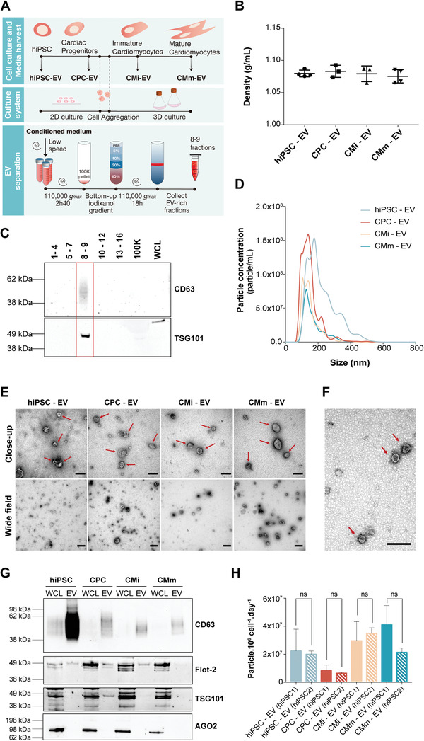
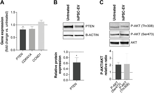
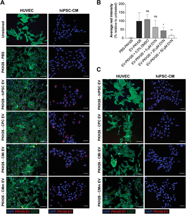
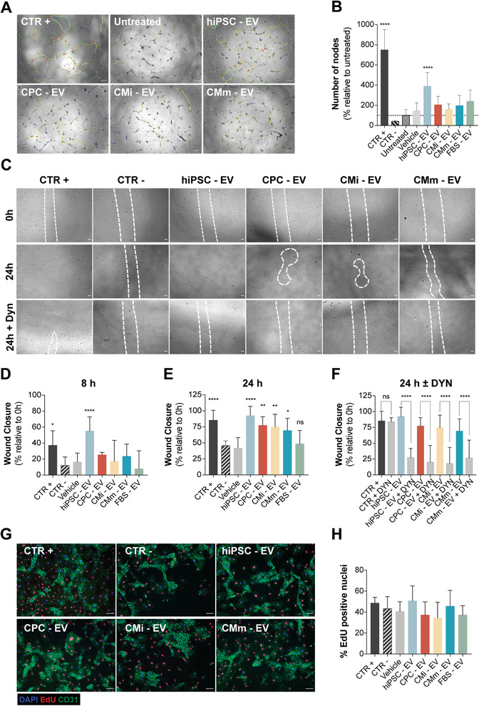
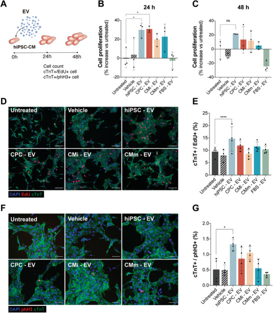
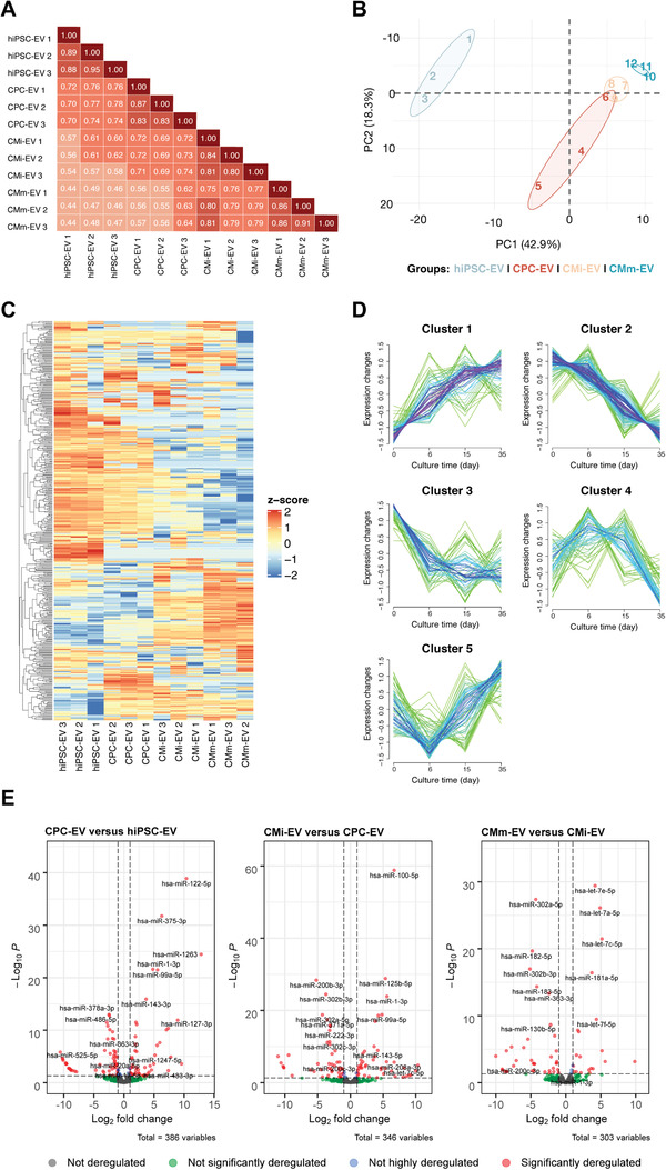
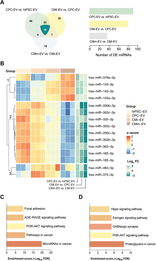
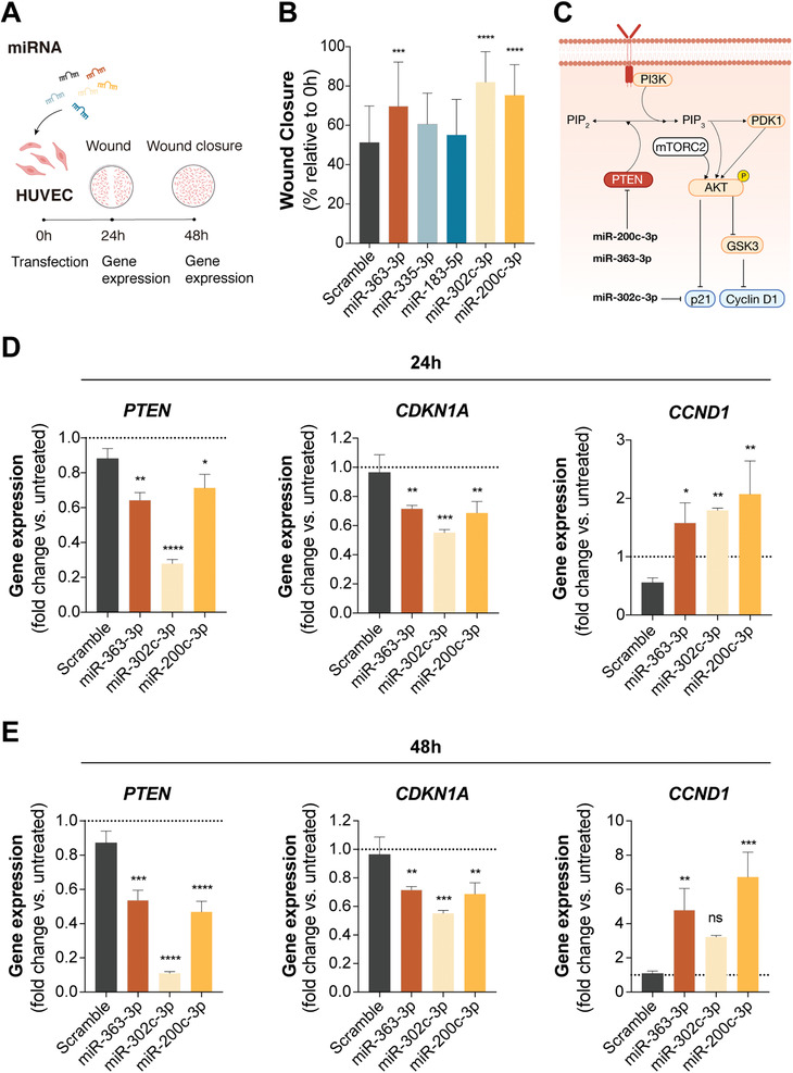
Extracellular vesicles (EV) are an attractive therapy to boost cardiac regeneration. Nevertheless, identification of native EV and corresponding cell platform(s) suitable for therapeutic application, is still a challenge. Here, EV are isolated from key stages of the human induced pluripotent stem cell-cardiomyocyte (hiPSC-CM) differentiation and maturation, i.e., from hiPSC (hiPSC-EV), cardiac progenitors, immature and mature cardiomyocytes, with the aim of identifying a promising cell biofactory for EV production, and pinpoint the genetic signatures of bioactive EV. EV secreted by hiPSC and cardiac derivatives show a typical size distribution profile and the expression of specific EV markers. Bioactivity assays show increased tube formation and migration in HUVEC treated with hiPSC-EV compared to EV from committed cell populations. hiPSC-EV also significantly increase cell cycle activity of hiPSC-CM. Global miRNA expression profiles, obtained by small RNA-seq analysis, corroborate an EV-miRNA pattern indicative of stem cell to cardiomyocyte specification, confirming that hiPSC-EV are enriched in pluripotency-associated miRNA with higher in vitro pro-angiogenic and pro-proliferative properties. In particular, a stemness maintenance miRNA cluster upregulated in hiPSC-EV targets the PTEN/PI3K/AKT pathway, involved in cell proliferation and survival. Overall, the findings validate hiPSC as cell biofactories for EV production for cardiac regenerative applications.

摘要

细胞外囊泡 (EV) 是一种很有吸引力的治疗方法,可以促进心脏再生。然而,鉴定天然 EV 及其适合治疗应用的相应细胞平台仍然是一个挑战。在这里,我们从人类诱导多能干细胞-心肌细胞 (hiPSC-CM) 分化和成熟的关键阶段分离 EV,即从 hiPSC (hiPSC-EV)、心脏祖细胞、未成熟和成熟的心肌细胞中分离 EV,目的是确定一个有前途的 EV 生产细胞工厂,并确定生物活性 EV 的遗传特征。hiPSC 和心脏衍生物分泌的 EV 显示出典型的大小分布特征和特定的 EV 标志物表达。生物活性测定显示,与来自定向细胞群的 EV 相比,用 hiPSC-EV 处理的 HUVEC 形成管和迁移的能力增加。hiPSC-EV 还显著增加 hiPSC-CM 的细胞周期活性。通过小 RNA-seq 分析获得的全球 miRNA 表达谱证实了 EV-miRNA 模式表明干细胞向心肌细胞特化,这证实了 hiPSC-EV 富含与体外更强的促血管生成和促增殖特性相关的多能性相关 miRNA。特别是,在 hiPSC-EV 中上调的一个维持干细胞特性的 miRNA 簇靶向参与细胞增殖和存活的 PTEN/PI3K/AKT 途径。总的来说,这些发现验证了 hiPSC 作为 EV 生产的细胞工厂,用于心脏再生应用。

https://cdn.ncbi.nlm.nih.gov/pmc/blobs/e110/9130911/65d233b5cc33/ADVS-9-2104296-g008.jpg
https://cdn.ncbi.nlm.nih.gov/pmc/blobs/e110/9130911/3265a193f3c0/ADVS-9-2104296-g001.jpg
https://cdn.ncbi.nlm.nih.gov/pmc/blobs/e110/9130911/1786b790e60d/ADVS-9-2104296-g006.jpg
https://cdn.ncbi.nlm.nih.gov/pmc/blobs/e110/9130911/649c715e686d/ADVS-9-2104296-g009.jpg
https://cdn.ncbi.nlm.nih.gov/pmc/blobs/e110/9130911/16aabd46ea51/ADVS-9-2104296-g002.jpg
https://cdn.ncbi.nlm.nih.gov/pmc/blobs/e110/9130911/ac45a498c239/ADVS-9-2104296-g003.jpg
https://cdn.ncbi.nlm.nih.gov/pmc/blobs/e110/9130911/e82799a8e384/ADVS-9-2104296-g005.jpg
https://cdn.ncbi.nlm.nih.gov/pmc/blobs/e110/9130911/433e113359e0/ADVS-9-2104296-g004.jpg
https://cdn.ncbi.nlm.nih.gov/pmc/blobs/e110/9130911/65d233b5cc33/ADVS-9-2104296-g008.jpg

相似文献

1
Bioactivity and miRNome Profiling of Native Extracellular Vesicles in Human Induced Pluripotent Stem Cell-Cardiomyocyte Differentiation.天然细胞外囊泡在人诱导多能干细胞向心肌细胞分化中的生物活性和 miRNA 谱分析。
Adv Sci (Weinh). 2022 May;9(15):e2104296. doi: 10.1002/advs.202104296. Epub 2022 Mar 24.
2
Expression of Extracellular Vesicle PIWI-Interacting RNAs Throughout hiPSC-Cardiomyocyte Differentiation.人诱导多能干细胞来源心肌细胞分化过程中细胞外囊泡PIWI相互作用RNA的表达
Front Physiol. 2022 Jun 16;13:926528. doi: 10.3389/fphys.2022.926528. eCollection 2022.
3
Extracellular vesicles from human cardiac stromal cells up-regulate cardiomyocyte protective responses to hypoxia.人心脏基质细胞来源的细胞外囊泡可上调心肌细胞对低氧的保护反应。
Stem Cell Res Ther. 2024 Oct 12;15(1):363. doi: 10.1186/s13287-024-03983-y.
4
Stem cell-derived extracellular vesicles reduce the expression of molecules involved in cardiac hypertrophy-In a model of human-induced pluripotent stem cell-derived cardiomyocytes.干细胞衍生的细胞外囊泡降低了参与心肌肥厚的分子的表达——在人诱导多能干细胞衍生的心肌细胞模型中。
Front Pharmacol. 2022 Oct 10;13:1003684. doi: 10.3389/fphar.2022.1003684. eCollection 2022.
5
Acellular therapeutic approach for heart failure: in vitro production of extracellular vesicles from human cardiovascular progenitors.细胞治疗心力衰竭的方法:从人心血管祖细胞体外生产细胞外囊泡。
Eur Heart J. 2018 May 21;39(20):1835-1847. doi: 10.1093/eurheartj/ehy012.
6
Extracellular Vesicles Released by Human Induced-Pluripotent Stem Cell-Derived Cardiomyocytes Promote Angiogenesis.人诱导多能干细胞衍生的心肌细胞释放的细胞外囊泡促进血管生成。
Front Physiol. 2018 Dec 14;9:1794. doi: 10.3389/fphys.2018.01794. eCollection 2018.
7
Time-regulated transcripts with the potential to modulate human pluripotent stem cell-derived cardiomyocyte differentiation.具有潜在调节人多能干细胞源性心肌细胞分化的时间调节转录物。
Stem Cell Res Ther. 2022 Sep 2;13(1):437. doi: 10.1186/s13287-022-03138-x.
8
Extracellular vesicles from human embryonic stem cell-derived cardiovascular progenitor cells promote cardiac infarct healing through reducing cardiomyocyte death and promoting angiogenesis.人胚胎干细胞来源的心血管祖细胞的细胞外囊泡通过减少心肌细胞死亡和促进血管生成促进心肌梗死愈合。
Cell Death Dis. 2020 May 11;11(5):354. doi: 10.1038/s41419-020-2508-y.
9
Differential Effects of Extracellular Vesicles of Lineage-Specific Human Pluripotent Stem Cells on the Cellular Behaviors of Isogenic Cortical Spheroids.谱系特异性人多能干细胞的细胞外囊泡对同源皮质类器官细胞行为的差异影响。
Cells. 2019 Aug 28;8(9):993. doi: 10.3390/cells8090993.
10
Donor-specific phenotypic variation in hiPSC cardiomyocyte-derived exosomes impacts endothelial cell function.供体特异性人诱导多能干细胞心肌细胞衍生外泌体表型变异影响血管内皮细胞功能。
Am J Physiol Heart Circ Physiol. 2021 Mar 1;320(3):H954-H968. doi: 10.1152/ajpheart.00463.2020. Epub 2021 Jan 8.

引用本文的文献

1
Extracellular Vesicles from Human Induced Pluripotent Stem Cells Exhibit a Unique MicroRNA and CircRNA Signature.来自人类诱导多能干细胞的细胞外囊泡呈现出独特的微小RNA和环状RNA特征。
Int J Biol Sci. 2024 Nov 22;20(15):6255-6278. doi: 10.7150/ijbs.100113. eCollection 2024.
2
Hypoxia and TNF-alpha modulate extracellular vesicle release from human induced pluripotent stem cell-derived cardiomyocytes.缺氧和 TNF-α调节人诱导多能干细胞源性心肌细胞释放细胞外囊泡。
J Extracell Vesicles. 2024 Nov;13(11):e70000. doi: 10.1002/jev2.70000.
3
A case for the study of native extracellular vesicles.

本文引用的文献

1
Assays and Imaging Methods for Drug Discovery for Cardiac Fibrosis.用于心脏纤维化药物发现的检测和成像方法
Front Physiol. 2021 Jul 8;12:697270. doi: 10.3389/fphys.2021.697270. eCollection 2021.
2
miR-106a-363 cluster in extracellular vesicles promotes endogenous myocardial repair via Notch3 pathway in ischemic heart injury.细胞外囊泡中的 miR-106a-363 簇通过 Notch3 通路促进缺血性心脏损伤中的内源性心肌修复。
Basic Res Cardiol. 2021 Mar 19;116(1):19. doi: 10.1007/s00395-021-00858-8.
3
Engineered models of the human heart: Directions and challenges.
一项关于天然细胞外囊泡的研究实例。
Front Oncol. 2024 Jul 12;14:1430971. doi: 10.3389/fonc.2024.1430971. eCollection 2024.
4
Hypoxic extracellular vesicles from hiPSCs protect cardiomyocytes from oxidative damage by transferring antioxidant proteins and enhancing Akt/Erk/NRF2 signaling.缺氧细胞外囊泡通过传递抗氧化蛋白和增强 Akt/Erk/NRF2 信号来保护心肌细胞免受氧化损伤。
Cell Commun Signal. 2024 Jul 9;22(1):356. doi: 10.1186/s12964-024-01722-7.
5
Extracellular vesicle biogenesis of three-dimensional human pluripotent stem cells in a novel Vertical-Wheel bioreactor.新型垂直轮生物反应器中三维人类多能干细胞的细胞外囊泡生物发生
J Extracell Biol. 2024 Jan 9;3(1):e133. doi: 10.1002/jex2.133. eCollection 2024 Jan.
6
Head-to-head comparison of relevant cell sources of small extracellular vesicles for cardiac repair: Superiority of embryonic stem cells.心脏修复中小细胞外囊泡相关细胞来源的头对头比较:胚胎干细胞更优。
J Extracell Vesicles. 2024 May;13(5):e12445. doi: 10.1002/jev2.12445.
7
m6A epitranscriptomic and epigenetic crosstalk in cardiac fibrosis.m6A 转录组和表观遗传学在心脏纤维化中的相互作用。
Mol Ther. 2024 Apr 3;32(4):878-889. doi: 10.1016/j.ymthe.2024.01.037. Epub 2024 Feb 3.
8
Extracellular vesicles derived from human ESC-MSCs target macrophage and promote anti-inflammation process, angiogenesis, and functional recovery in ACS-induced severe skeletal muscle injury.人胚胎干细胞来源的细胞外囊泡靶向巨噬细胞,促进 ACS 诱导的严重骨骼肌损伤中的抗炎过程、血管生成和功能恢复。
Stem Cell Res Ther. 2023 Nov 14;14(1):331. doi: 10.1186/s13287-023-03530-1.
9
Clinically Expired Platelet Concentrates as a Source of Extracellular Vesicles for Targeted Anti-Cancer Drug Delivery.临床过期血小板浓缩物作为用于靶向抗癌药物递送的细胞外囊泡来源
Pharmaceutics. 2023 Mar 15;15(3):953. doi: 10.3390/pharmaceutics15030953.
10
Electric-Field-Driven Printed 3D Highly Ordered Microstructure with Cell Feature Size Promotes the Maturation of Engineered Cardiac Tissues.电场驱动打印 3D 高度有序微结构具有细胞特征尺寸促进工程心脏组织的成熟。
Adv Sci (Weinh). 2023 Apr;10(11):e2206264. doi: 10.1002/advs.202206264. Epub 2023 Feb 13.
人工心脏模型:方向与挑战。
Stem Cell Reports. 2021 Sep 14;16(9):2049-2057. doi: 10.1016/j.stemcr.2020.11.013. Epub 2020 Dec 18.
4
miR-301a-PTEN-AKT Signaling Induces Cardiomyocyte Proliferation and Promotes Cardiac Repair Post-MI.miR-301a-PTEN-AKT信号通路诱导心肌细胞增殖并促进心肌梗死后的心脏修复。
Mol Ther Nucleic Acids. 2020 Aug 29;22:251-262. doi: 10.1016/j.omtn.2020.08.033. eCollection 2020 Dec 4.
5
Cells of the adult human heart.成人心脏细胞。
Nature. 2020 Dec;588(7838):466-472. doi: 10.1038/s41586-020-2797-4. Epub 2020 Sep 24.
6
Exosomes secreted by hiPSC-derived cardiac cells improve recovery from myocardial infarction in swine.人诱导多能干细胞衍生的心肌细胞分泌的细胞外囊泡可改善猪心肌梗死后的恢复。
Sci Transl Med. 2020 Sep 16;12(561). doi: 10.1126/scitranslmed.aay1318.
7
Forced expression of miR-143 and -145 in cardiomyocytes induces cardiomyopathy with a reductive redox shift.在心肌细胞中强制表达 miR-143 和 -145 会导致氧化还原状态降低的心肌病。
Cell Mol Biol Lett. 2020 Aug 24;25:40. doi: 10.1186/s11658-020-00232-x. eCollection 2020.
8
Extracellular Vesicles as Therapeutic Agents for Cardiac Fibrosis.细胞外囊泡作为心脏纤维化的治疗剂
Front Physiol. 2020 May 21;11:479. doi: 10.3389/fphys.2020.00479. eCollection 2020.
9
CDC-derived extracellular vesicles reprogram inflammatory macrophages to an arginase 1-dependent proangiogenic phenotype.CDC 衍生的细胞外囊泡将炎症巨噬细胞重编程为依赖精氨酸酶 1 的促血管生成表型。
Am J Physiol Heart Circ Physiol. 2020 Jun 1;318(6):H1447-H1460. doi: 10.1152/ajpheart.00155.2020. Epub 2020 Apr 24.
10
Extracellular vesicles from human cardiovascular progenitors trigger a reparative immune response in infarcted hearts.来自人类心血管祖细胞的细胞外囊泡在梗死心脏中引发修复性免疫反应。
Cardiovasc Res. 2021 Jan 1;117(1):292-307. doi: 10.1093/cvr/cvaa028.