Komatsu Shugo, Terui Keita, Nakata Mitsuyuki, Shibata Ryohei, Oita Satoru, Kawaguchi Yunosuke, Yoshizawa Hiroko, Hirokawa Tomoya, Nakatani Erika, Hishiki Tomoro
Department of Pediatric Surgery, Chiba University Graduate School of Medicine, 1-8-1 Inohana, Chuo-ku, Chiba 260-8677, Japan.
Children (Basel). 2022 Mar 8;9(3):376. doi: 10.3390/children9030376.
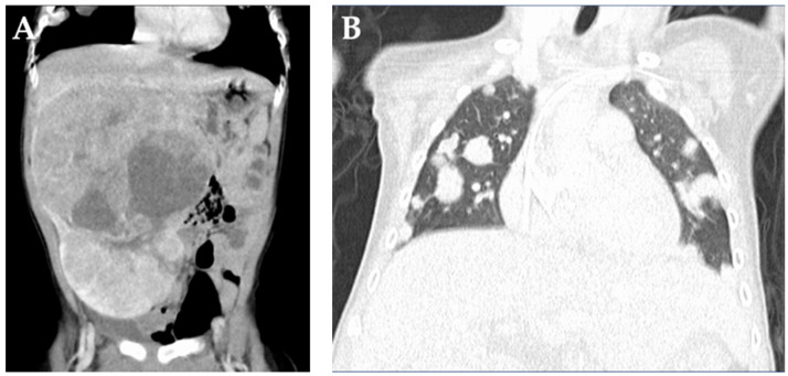
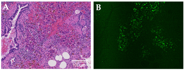
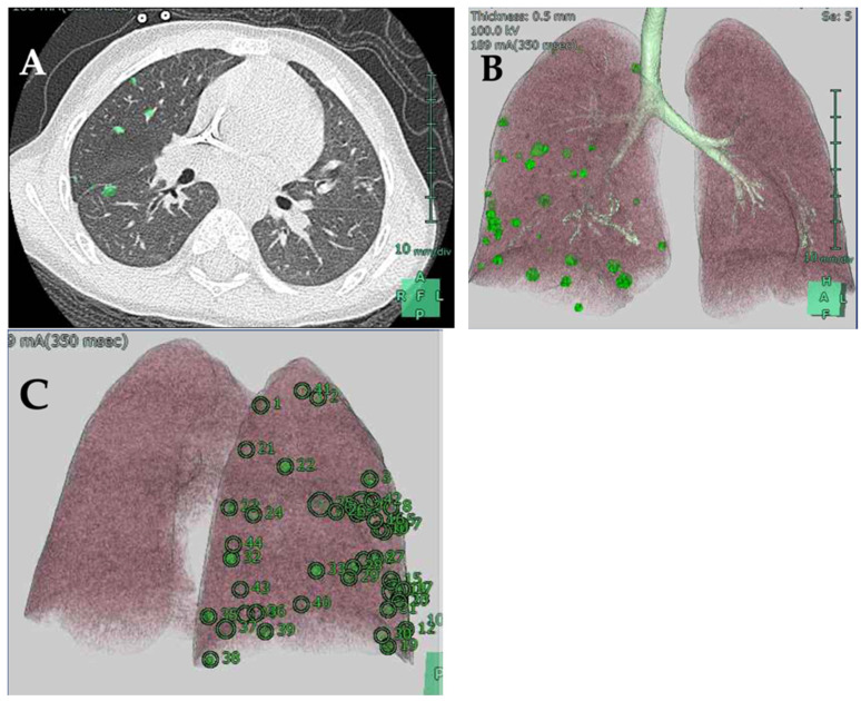
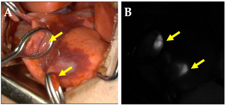
It is essential to accurately and safely resect all tumors during surgery for multiple lung metastases. Here, we report a case of hepatoblastoma (HB) with multiple pulmonary nodules that ultimately underwent complete resection using combined three-dimensional image reconstruction and indocyanine green (ICG) fluorescence guidance. A 1-year-old boy was diagnosed with HB and multiple lung metastases. After intensive chemotherapy, complete resection with subsegmentectomy (S5 + 6) and partial resection (S3, S8) were performed. More than 100 pulmonary nodules, which remained visible on computed tomography (CT) despite additional postoperative chemotherapy, were subjected to pulmonary resection. We used the SYNAPSE VINCENT software (Fujifilm Medical, Tokyo, Japan) to obtain three-dimensional images of the nodules. We numbered each nodule, and 33 lesions of the right lung were resected by multiple wedge resections through a right thoracotomy, with the aid of palpation and ICG fluorescence guidance. One month after the right metastasectomy, resection of 64 lesions in the left lung was performed via left thoracotomy. Postoperative CT showed complete clearance of the lung lesions, and the patient remained disease-free for 15 months after the treatment. This case study confirms that the combination of three-dimensional localization and ICG fluorescence guidance allows for accurate and safe resection of nearly 100 lung metastases.
在手术治疗多发性肺转移瘤时,准确且安全地切除所有肿瘤至关重要。在此,我们报告一例肝母细胞瘤(HB)合并多个肺结节的病例,该病例最终通过三维图像重建和吲哚菁绿(ICG)荧光引导实现了完全切除。一名1岁男孩被诊断为HB并伴有多个肺转移瘤。经过强化化疗后,进行了亚段切除术(S5 + 6)和部分切除术(S3、S8)。尽管术后进行了额外化疗,但计算机断层扫描(CT)上仍可见100多个肺结节,遂对这些结节进行了肺切除。我们使用SYNAPSE VINCENT软件(富士胶片医疗,日本东京)获取结节的三维图像。我们对每个结节进行编号,并通过右胸切开术借助触诊和ICG荧光引导,经多次楔形切除术切除了右肺的33个病灶。右肺转移瘤切除术后1个月,通过左胸切开术对左肺的64个病灶进行了切除。术后CT显示肺部病灶完全清除,治疗后患者无病生存15个月。本病例研究证实,三维定位与ICG荧光引导相结合可实现近100个肺转移瘤的准确、安全切除。