Bundschuh Lena, Prokic Vesna, Guckenberger Matthias, Tanadini-Lang Stephanie, Essler Markus, Bundschuh Ralph A
Department of Nuclear Medicine, University Hospital Bonn, 53127 Bonn, Germany.
Department of Physics, University Koblenz-Landau, 55118 Koblenz, Germany.
Diagnostics (Basel). 2022 Feb 23;12(3):576. doi: 10.3390/diagnostics12030576.
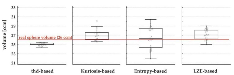
Positron emission tomography (PET) provides important additional information when applied in radiation therapy treatment planning. However, the optimal way to define tumors in PET images is still undetermined. As radiomics features are gaining more and more importance in PET image interpretation as well, we aimed to use textural features for an optimal differentiation between tumoral tissue and surrounding tissue to segment-target lesions based on three textural parameters found to be suitable in previous analysis (Kurtosis, Local Entropy and Long Zone Emphasis). Intended for use in radiation therapy planning, this algorithm was combined with a previously described motion-correction algorithm and validated in phantom data. In addition, feasibility was shown in five patients. The algorithms provided sufficient results for phantom and patient data. The stability of the results was analyzed in 20 consecutive measurements of phantom data. Results for textural feature-based algorithms were slightly worse than those of the threshold-based reference algorithm (mean standard deviation 1.2%-compared to 4.2% to 8.6%) However, the Entropy-based algorithm came the closest to the real volume of the phantom sphere of 6 ccm with a mean measured volume of 26.5 ccm. The threshold-based algorithm found a mean volume of 25.0 ccm. In conclusion, we showed a novel, radiomics-based tumor segmentation algorithm in FDG-PET with promising results in phantom studies concerning recovered lesion volume and reasonable results in stability in consecutive measurements. Segmentation based on Entropy was the most precise in comparison with sphere volume but showed the worst stability in consecutive measurements. Despite these promising results, further studies with larger patient cohorts and histopathological standards need to be performed for further validation of the presented algorithms and their applicability in clinical routines. In addition, their application in other tumor entities needs to be studied.
正电子发射断层扫描(PET)应用于放射治疗治疗计划时可提供重要的额外信息。然而,在PET图像中定义肿瘤的最佳方法仍未确定。由于放射组学特征在PET图像解读中也越来越重要,我们旨在基于先前分析中发现合适的三个纹理参数(峰度、局部熵和长区域强调),使用纹理特征对肿瘤组织和周围组织进行最佳区分,以分割目标病变。该算法旨在用于放射治疗计划,与先前描述的运动校正算法相结合,并在体模数据中进行了验证。此外,在五名患者中展示了其可行性。这些算法为体模和患者数据提供了充分的结果。对体模数据进行了连续20次测量以分析结果的稳定性。基于纹理特征的算法的结果略逊于基于阈值的参考算法(平均标准差为1.2%,而基于阈值的算法为4.2%至8.6%)。然而,基于熵的算法最接近6立方厘米体模球体的实际体积,平均测量体积为26.5立方厘米。基于阈值的算法测得的平均体积为25.0立方厘米。总之,我们展示了一种基于放射组学的新型FDG-PET肿瘤分割算法,在体模研究中关于恢复病变体积方面有很有前景的结果,在连续测量的稳定性方面有合理的结果。与球体体积相比,基于熵的分割最为精确,但在连续测量中稳定性最差。尽管有这些有前景的结果,但仍需要对更大的患者队列进行进一步研究,并采用组织病理学标准,以进一步验证所提出的算法及其在临床常规中的适用性。此外,还需要研究它们在其他肿瘤实体中的应用。