Mautuit Thibaud, Semecas Rachel, Hogg Stephen, Daien Vincent, Gavard Olivier, Chateau Nicolas, MacGillivray Tom, Trucco Emanuele, Chiquet Christophe
Department of Ophthalmology, University Hospital of Grenoble-Alpes, University Grenoble Alpes, Inserm, CHU Grenoble Alpes, HP2, 38000 Grenoble, France.
VAMPIRE Project, Computing (SSE), University of Dundee, Dundee DD1 4HN, UK.
Diagnostics (Basel). 2022 Mar 13;12(3):705. doi: 10.3390/diagnostics12030705.
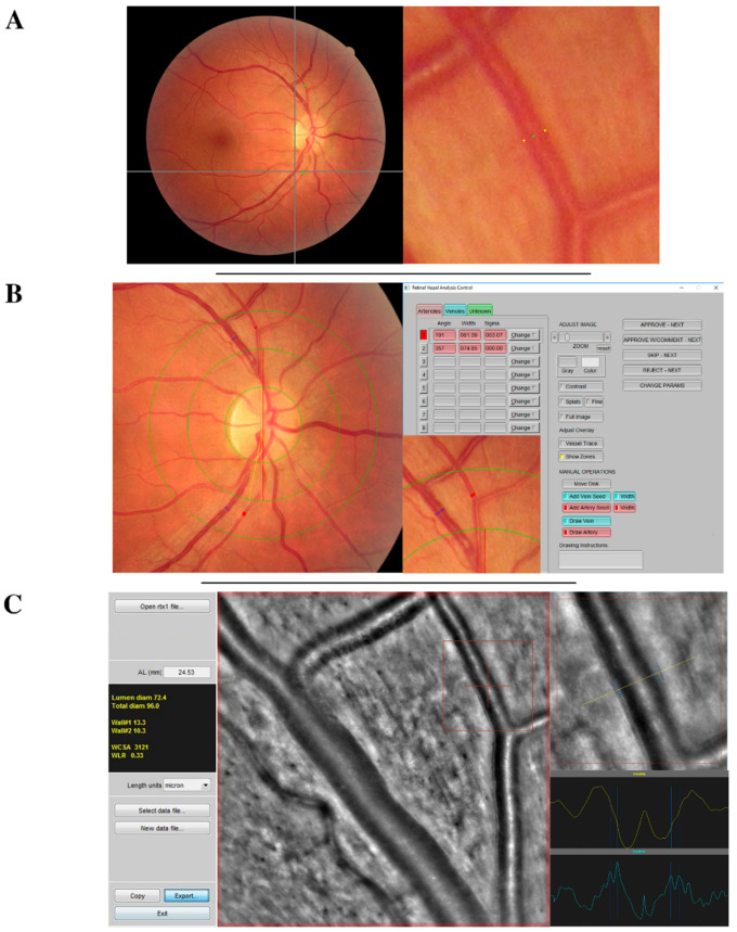
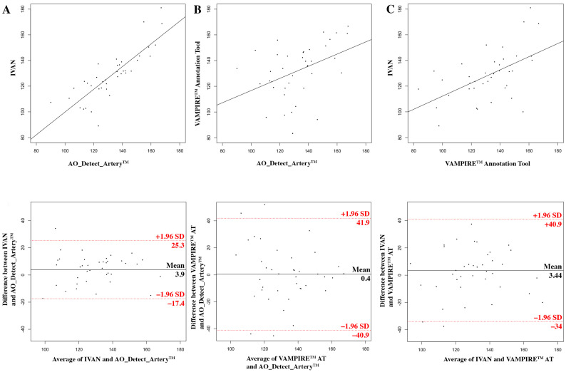
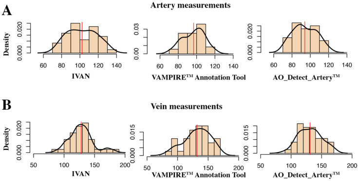
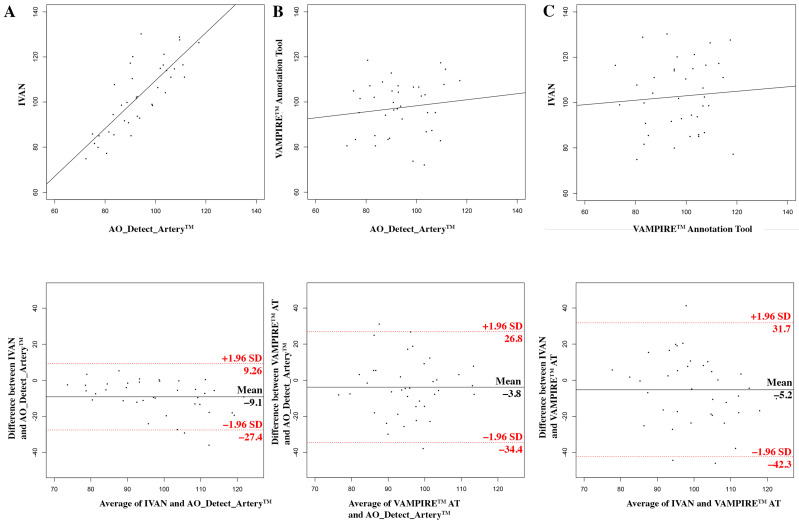
The aim of this prospective study was to compare retinal vascular diameter measurements taken from standard fundus images and adaptive optics (AO) images. We analysed retinal images of twenty healthy subjects with 45-degree funduscopic colour photographs (CR-2 Canon fundus camera, Canon™) and adaptive optics (AO) fundus images (rtx1 camera, Imagine Eyes). Diameters were measured using three software applications: the VAMPIRE (Vessel Assessment and Measurement Platform for Images of the REtina) annotation tool, IVAN (Interactive Vessel ANalyzer) for funduscopic colour photographs, and AO_Detect_Artery™ for AO images. For the arterial diameters, the mean difference between AO_Detect_Artery™ and IVAN was 9.1 µm (-27.4 to 9.2 µm, = 0.005) and the measurements were significantly correlated (r = 0.79). The mean difference between AO_Detect_Artery™ and VAMPIRE annotation tool was 3.8 µm (-34.4 to 26.8 µm, = 0.16) and the measurements were poorly correlated (r = 0.12). For the venous diameters, the mean difference between the AO_Detect_Artery™ and IVAN was 3.9 µm (-40.9 to 41.9 µm, = 0.35) and the measurements were highly correlated (r = 0.83). The mean difference between the AO_Detect_Artery™ and VAMPIRE annotation tool was 0.4 µm (-17.44 to 25.3 µm, = 0.91) and the correlations were moderate (r = 0.41). We found that the VAMPIRE annotation tool, an entirely manual software, is accurate for the measurement of arterial and venular diameters, but the correlation with AO measurements is poor. On the contrary, IVAN, a semi-automatic software tool, presents slightly greater differences with AO imaging, but the correlation is stronger. Data from arteries should be considered with caution, since IVAN seems to significantly under-estimate arterial diameters.
这项前瞻性研究的目的是比较从标准眼底图像和自适应光学(AO)图像获取的视网膜血管直径测量值。我们分析了20名健康受试者的视网膜图像,包括用45度眼底彩色照片(CR - 2佳能眼底相机,佳能公司)和自适应光学(AO)眼底图像(rtx1相机,Imagine Eyes公司)。使用三种软件应用程序测量直径:用于视网膜图像的血管评估和测量平台(VAMPIRE)标注工具、用于眼底彩色照片的交互式血管分析仪(IVAN)以及用于AO图像的AO_Detect_Artery™。对于动脉直径,AO_Detect_Artery™与IVAN之间的平均差异为9.1微米(-27.4至9.2微米,P = 0.005),且测量值显著相关(r = 0.79)。AO_Detect_Artery™与VAMPIRE标注工具之间的平均差异为3.8微米(-34.4至26.8微米,P = 0.16),且测量值相关性较差(r = 0.12)。对于静脉直径,AO_Detect_Artery™与IVAN之间的平均差异为3.9微米(-40.9至41.9微米,P = 0.35),且测量值高度相关(r = 0.83)。AO_Detect_Artery™与VAMPIRE标注工具之间的平均差异为0.4微米(-17.44至25.3微米,P = 0.91),且相关性中等(r = 0.41)。我们发现,VAMPIRE标注工具,一个完全手动的软件,在测量动脉和静脉直径方面是准确的,但与AO测量的相关性较差。相反,IVAN,一个半自动软件工具,与AO成像的差异稍大,但相关性更强。由于IVAN似乎显著低估动脉直径,动脉数据应谨慎考虑。