• 文献检索
  • 文档翻译
  • 深度研究
  • 学术资讯
  • Suppr Zotero 插件Zotero 插件
  • 邀请有礼
  • 套餐&价格
  • 历史记录
应用&插件
Suppr Zotero 插件Zotero 插件浏览器插件Mac 客户端Windows 客户端微信小程序
定价
高级版会员购买积分包购买API积分包
服务
文献检索文档翻译深度研究API 文档MCP 服务
关于我们
关于 Suppr公司介绍联系我们用户协议隐私条款
关注我们

Suppr 超能文献

核心技术专利:CN118964589B侵权必究
粤ICP备2023148730 号-1Suppr @ 2026

文献检索

告别复杂PubMed语法,用中文像聊天一样搜索,搜遍4000万医学文献。AI智能推荐,让科研检索更轻松。

立即免费搜索

文件翻译

保留排版,准确专业,支持PDF/Word/PPT等文件格式,支持 12+语言互译。

免费翻译文档

深度研究

AI帮你快速写综述,25分钟生成高质量综述,智能提取关键信息,辅助科研写作。

立即免费体验

碘酸钠注射液可复制色素性小鼠和大鼠的视网膜退行性病变阶段:使用 OCT 和 ERG 进行非侵入性随访。

Sodium-Iodate Injection Can Replicate Retinal Degenerative Disease Stages in Pigmented Mice and Rats: Non-Invasive Follow-Up Using OCT and ERG.

机构信息

Department of Human Genetics, Section Ophthalmogenetics, Amsterdam University Medical Centers (AUMC), University of Amsterdam (UvA), Location AMC, Meibergdreef, 1105 AZ Amsterdam, The Netherlands.

Georgia Institute of Technology, G.W. Woodruff School of Mechanical Engineering, Atlanta, GA 30332, USA.

出版信息

Int J Mol Sci. 2022 Mar 8;23(6):2918. doi: 10.3390/ijms23062918.

DOI:10.3390/ijms23062918
PMID:35328338
原文链接:https://pmc.ncbi.nlm.nih.gov/articles/PMC8953416/
Abstract

PURPOSE

The lack of suitable animal models for (dry) age-related macular degeneration (AMD) has hampered therapeutic research into the disease, so far. In this study, pigmented rats and mice were systematically injected with various doses of sodium iodate (SI). After injection, the retinal structure and visual function were non-invasively characterized over time to obtain in-depth data on the suitability of these models for studying experimental therapies for retinal degenerative diseases, such as dry AMD.

METHODS

SI was injected into the tail vein (i.v.) using a series of doses (0-70 mg/kg) in adolescent C57BL/6J mice and Brown Norway rats. The retinal structure and function were assessed non-invasively at baseline (day 1) and at several time points (1-3, 5, and 10-weeks) post-injection by scanning laser ophthalmoscopy (SLO), optical coherence tomography (OCT), and electroretinography (ERG).

RESULTS

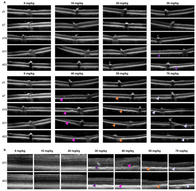
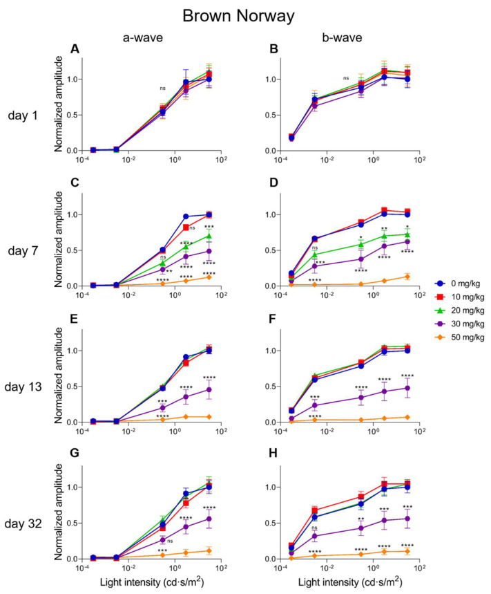
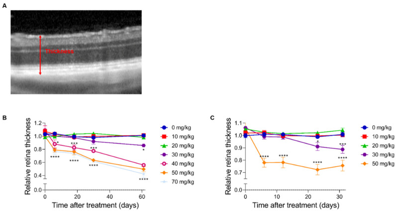
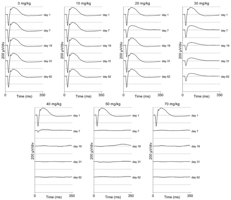
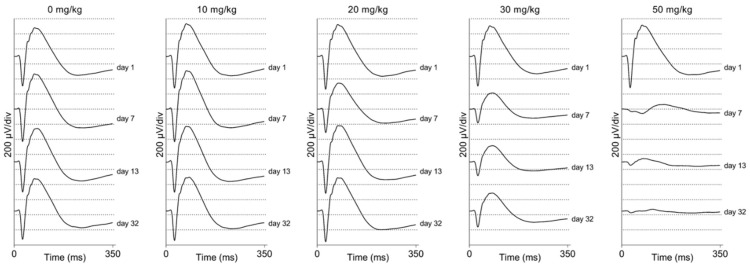
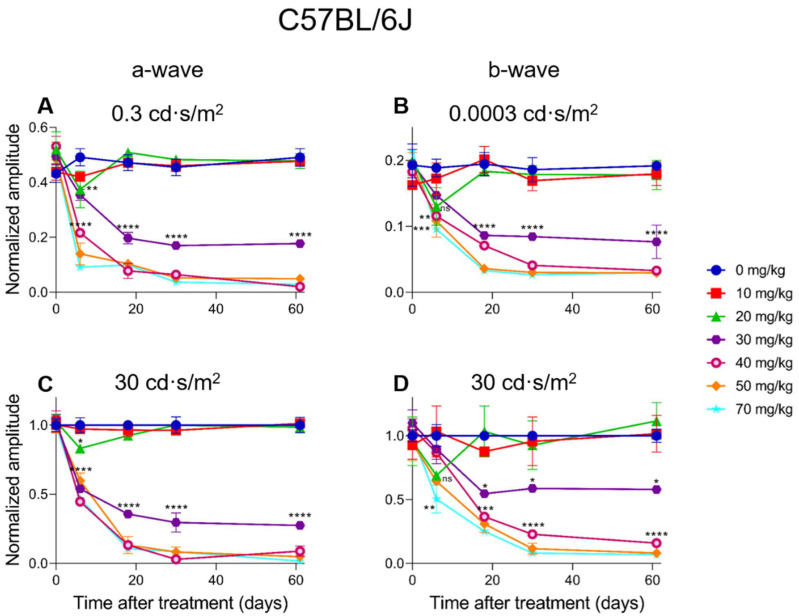
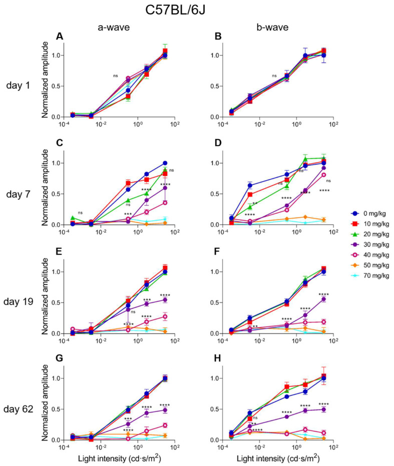
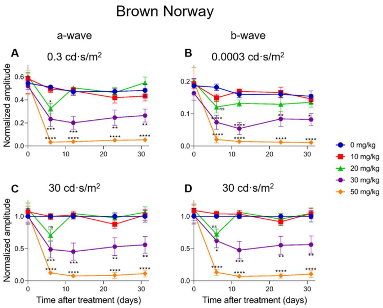
After the SI injection, retinal degeneration in mice and rats yielded similar results. The lowest dose (10 mg/kg) resulted in non-detectable structural or functional effects. An injection with 20 mg/kg SI did not result in an evident retinal degeneration as judged from the OCT data. In contrast, the ERG responses were temporarily decreased but returned to baseline within two-weeks. Higher doses (30, 40, 50, and 70 mg/kg) resulted in moderate to severe structural RPE and retinal injury and decreased the ERG amplitudes, indicating visual impairment in both mice and rat strains.

CONCLUSIONS

After the SI injections, we observed dose-dependent structural and functional pathological effects on the retinal pigment epithelium (RPE) and retina in the pigmented mouse and rat strains that were used in this study. Similar effects were observed in both species. In particular, a dose of 30 mg/kg seems to be suitable for future studies on developing experimental therapies. These relatively easily induced non-inherited models may serve as useful tools for evaluating novel therapies for RPE-related retinal degenerations, such as AMD.

摘要

目的

缺乏合适的动物模型一直阻碍着与年龄相关的干性黄斑变性(AMD)相关的治疗研究。在这项研究中,我们系统性地给色素沉着的大鼠和小鼠尾静脉注射不同剂量的碘酸钠(SI)。注射后,我们通过扫频激光检眼镜(SLO)、光学相干断层扫描(OCT)和视网膜电图(ERG)等非侵入性方法,随时间对视网膜结构和视觉功能进行了特征描述,以深入了解这些模型是否适合研究干性 AMD 等视网膜退行性疾病的实验性治疗方法。

方法

我们用一系列剂量(0-70mg/kg)在青春期 C57BL/6J 小鼠和棕色挪威鼠的尾静脉(i.v.)中注射 SI。在基线(第 1 天)和注射后 1-3、5 和 10 周的多个时间点,我们通过 SLO、OCT 和 ERG 等非侵入性方法评估视网膜结构和功能。

结果

SI 注射后,小鼠和大鼠的视网膜变性产生了相似的结果。最低剂量(10mg/kg)导致结构或功能无明显变化。从 OCT 数据来看,20mg/kg SI 注射不会导致明显的视网膜变性。相反,ERG 反应暂时降低,但在两周内恢复到基线。更高剂量(30、40、50 和 70mg/kg)导致中重度 RPE 和视网膜损伤,以及 ERG 幅度降低,表明两种品系的小鼠和大鼠都出现了视力障碍。

结论

在 SI 注射后,我们观察到在用于本研究的色素沉着的小鼠和大鼠品系的视网膜色素上皮(RPE)和视网膜上出现了剂量依赖性的结构和功能病理变化。两种物种都观察到了相似的效果。特别是,30mg/kg 的剂量似乎适合用于开发实验性治疗方法的未来研究。这些相对容易诱导的非遗传性模型可能成为评估 RPE 相关视网膜变性(如 AMD)新型治疗方法的有用工具。

https://cdn.ncbi.nlm.nih.gov/pmc/blobs/951c/8953416/c8e484bd5f9c/ijms-23-02918-g009.jpg
https://cdn.ncbi.nlm.nih.gov/pmc/blobs/951c/8953416/f26a100f54c0/ijms-23-02918-g001.jpg
https://cdn.ncbi.nlm.nih.gov/pmc/blobs/951c/8953416/2c950e4c8250/ijms-23-02918-g002.jpg
https://cdn.ncbi.nlm.nih.gov/pmc/blobs/951c/8953416/1e6535a18b71/ijms-23-02918-g003.jpg
https://cdn.ncbi.nlm.nih.gov/pmc/blobs/951c/8953416/f566cae0dbbb/ijms-23-02918-g004.jpg
https://cdn.ncbi.nlm.nih.gov/pmc/blobs/951c/8953416/bde485412b7e/ijms-23-02918-g005.jpg
https://cdn.ncbi.nlm.nih.gov/pmc/blobs/951c/8953416/8c33f1657750/ijms-23-02918-g006.jpg
https://cdn.ncbi.nlm.nih.gov/pmc/blobs/951c/8953416/647351704658/ijms-23-02918-g007.jpg
https://cdn.ncbi.nlm.nih.gov/pmc/blobs/951c/8953416/5d4087db389d/ijms-23-02918-g008.jpg
https://cdn.ncbi.nlm.nih.gov/pmc/blobs/951c/8953416/c8e484bd5f9c/ijms-23-02918-g009.jpg

相似文献

1
Sodium-Iodate Injection Can Replicate Retinal Degenerative Disease Stages in Pigmented Mice and Rats: Non-Invasive Follow-Up Using OCT and ERG.碘酸钠注射液可复制色素性小鼠和大鼠的视网膜退行性病变阶段:使用 OCT 和 ERG 进行非侵入性随访。
Int J Mol Sci. 2022 Mar 8;23(6):2918. doi: 10.3390/ijms23062918.
2
Course of Sodium Iodate-Induced Retinal Degeneration in Albino and Pigmented Mice.白化小鼠和有色小鼠中碘酸钠诱导的视网膜变性病程
Invest Ophthalmol Vis Sci. 2017 Apr 1;58(4):2239-2249. doi: 10.1167/iovs.16-21255.
3
Irreversible Photoreceptors and RPE Cells Damage by Intravenous Sodium Iodate in Mice Is Related to Macrophage Accumulation.静脉注射碘酸钠导致小鼠光感受器和 RPE 细胞不可逆转的损伤与巨噬细胞的积累有关。
Invest Ophthalmol Vis Sci. 2018 Jul 2;59(8):3476-3487. doi: 10.1167/iovs.17-23532.
4
Inhibition of HDAC1 and 3 in the Presence of Systemic Inflammation Reduces Retinal Degeneration in a Model of Dry Age-Related Macular Degeneration.在系统性炎症存在的情况下抑制 HDAC1 和 3 可减少干性年龄相关性黄斑变性模型中的视网膜变性。
J Ocul Pharmacol Ther. 2024 Jul-Aug;40(6):397-406. doi: 10.1089/jop.2023.0163. Epub 2024 Apr 12.
5
Inducible RPE-specific GPX4 knockout causes oxidative stress and retinal degeneration with features of age-related macular degeneration.诱导型 RPE 特异性 GPX4 敲除导致氧化应激和视网膜变性,具有年龄相关性黄斑变性的特征。
Exp Eye Res. 2024 Oct;247:110028. doi: 10.1016/j.exer.2024.110028. Epub 2024 Aug 10.
6
Sodium-iodate injection can replicate retinal and choroid degeneration in pigmented mice: Using multimodal imaging and label-free quantitative proteomics analysis.碘酸钠注射液可复制色素性小鼠的视网膜和脉络膜变性:应用多模态成像和无标记定量蛋白质组学分析。
Exp Eye Res. 2024 Oct;247:110050. doi: 10.1016/j.exer.2024.110050. Epub 2024 Aug 14.
7
Tamoxifen protects photoreceptors in the sodium iodate model.他莫昔芬可保护碘酸钠模型中的光感受器。
Exp Eye Res. 2024 May;242:109879. doi: 10.1016/j.exer.2024.109879. Epub 2024 Apr 1.
8
Optimizing the sodium iodate model: Effects of dose, gender, and age.优化碘酸钠模型:剂量、性别和年龄的影响。
Exp Eye Res. 2024 Feb;239:109772. doi: 10.1016/j.exer.2023.109772. Epub 2023 Dec 27.
9
Retinal degeneration rat model: A study on the structural and functional changes in the retina following injection of sodium iodate.视网膜变性大鼠模型:碘酸钠注射后视网膜结构和功能变化的研究。
J Photochem Photobiol B. 2019 Jul;196:111514. doi: 10.1016/j.jphotobiol.2019.111514. Epub 2019 May 22.
10
The effects of intravitreal sodium iodate injection on retinal degeneration following vitrectomy in rabbits.玻璃体内注射碘酸钠对兔玻璃体切割术后视网膜变性的影响。
Sci Rep. 2019 Oct 30;9(1):15696. doi: 10.1038/s41598-019-52172-y.

引用本文的文献

1
Evaluation of retinal structure changes with AI-based OCT image segmentation for sodium iodate induced retinal degeneration.基于人工智能的光学相干断层扫描(OCT)图像分割技术评估碘酸钠诱导的视网膜变性中的视网膜结构变化
Front Cell Neurosci. 2025 Jun 18;19:1605639. doi: 10.3389/fncel.2025.1605639. eCollection 2025.
2
Single-cell transcriptome atlas of spontaneous dry age-related macular degeneration in macaques.猕猴自发性干性年龄相关性黄斑变性的单细胞转录组图谱
Fundam Res. 2023 Apr 25;5(3):1034-1046. doi: 10.1016/j.fmre.2023.02.028. eCollection 2025 May.
3
Animal models for the evaluation of retinal stem cell therapies.

本文引用的文献

1
Epithelial phenotype restoring drugs suppress macular degeneration phenotypes in an iPSC model.上皮表型恢复药物抑制 iPSC 模型中的黄斑变性表型。
Nat Commun. 2021 Dec 15;12(1):7293. doi: 10.1038/s41467-021-27488-x.
2
The Lrat Rat: CRISPR/Cas9 Construction and Phenotyping of a New Animal Model for Retinitis Pigmentosa.Lrat 大鼠:用于色素性视网膜炎的新型动物模型的 CRISPR/Cas9 构建和表型分析。
Int J Mol Sci. 2021 Jul 5;22(13):7234. doi: 10.3390/ijms22137234.
3
The interplay of oxidative stress and ARMS2-HTRA1 genetic risk in neovascular AMD.
用于评估视网膜干细胞疗法的动物模型。
Prog Retin Eye Res. 2025 May;106:101356. doi: 10.1016/j.preteyeres.2025.101356. Epub 2025 Apr 14.
4
Retinal Protective Effect of Mono-Ethyl Fumarate in Experimental Age-Related Macular Degeneration via Anti-Oxidative and Anti-Apoptotic Alterations.富马酸单乙酯通过抗氧化和抗凋亡改变对实验性年龄相关性黄斑变性的视网膜保护作用
Int J Mol Sci. 2025 Feb 7;26(4):1413. doi: 10.3390/ijms26041413.
5
Potential Use of Plasma Rich in Growth Factors in Age-Related Macular Degeneration: Evidence from a Mouse Model.富含生长因子的血浆在年龄相关性黄斑变性中的潜在应用:来自小鼠模型的证据。
Medicina (Kaunas). 2024 Dec 10;60(12):2036. doi: 10.3390/medicina60122036.
6
Deletion of the stress response protein REDD1 prevents sodium iodate-induced RPE damage and photoreceptor loss.应激反应蛋白REDD1的缺失可预防碘酸钠诱导的视网膜色素上皮(RPE)损伤和光感受器丧失。
Geroscience. 2025 Apr;47(2):1789-1803. doi: 10.1007/s11357-024-01362-2. Epub 2024 Oct 5.
7
Protective Effect and Related Mechanism of Modified Danggui Buxue Decoction on Retinal Oxidative Damage in Mice based on Network Pharmacology.基于网络药理学的加味当归补血汤对小鼠视网膜氧化损伤的保护作用及相关机制。
Curr Pharm Des. 2024;30(24):1912-1926. doi: 10.2174/0113816128293824240517113238.
8
Multi-Wavelength Photobiomodulation Ameliorates Sodium Iodate-Induced Age-Related Macular Degeneration in Rats.多波长光生物调节改善碘酸钠诱导的大鼠年龄相关性黄斑变性。
Int J Mol Sci. 2023 Dec 12;24(24):17394. doi: 10.3390/ijms242417394.
9
Regulated cell death pathways in the sodium iodate model: Insights and implications for AMD.碘酸钠模型中的调控细胞死亡途径:对 AMD 的启示和影响。
Exp Eye Res. 2024 Jan;238:109728. doi: 10.1016/j.exer.2023.109728. Epub 2023 Nov 14.
10
Paraoxonase 2 Deficiency Causes Mitochondrial Dysfunction in Retinal Pigment Epithelial Cells and Retinal Degeneration in Mice.对氧磷酶2缺乏导致小鼠视网膜色素上皮细胞线粒体功能障碍和视网膜退化。
Antioxidants (Basel). 2023 Sep 30;12(10):1820. doi: 10.3390/antiox12101820.
氧化应激与ARMS2-HTRA1基因风险在新生血管性年龄相关性黄斑变性中的相互作用。
Vessel Plus. 2021;5. doi: 10.20517/2574-1209.2020.48. Epub 2021 Jan 15.
4
Age-related macular degeneration.年龄相关性黄斑变性。
Nat Rev Dis Primers. 2021 May 6;7(1):31. doi: 10.1038/s41572-021-00265-2.
5
AMD Genetics: Methods and Analyses for Association, Progression, and Prediction.AMD 遗传学:关联、进展和预测的方法与分析。
Adv Exp Med Biol. 2021;1256:191-200. doi: 10.1007/978-3-030-66014-7_7.
6
The Impact of Oxidative Stress on Blood-Retinal Barrier Physiology in Age-Related Macular Degeneration.氧化应激对年龄相关性黄斑变性血视网膜屏障生理学的影响。
Cells. 2021 Jan 4;10(1):64. doi: 10.3390/cells10010064.
7
Progress in developing rodent models of age-related macular degeneration (AMD).与年龄相关的黄斑变性(AMD)的啮齿动物模型的研究进展。
Exp Eye Res. 2021 Feb;203:108404. doi: 10.1016/j.exer.2020.108404. Epub 2020 Dec 17.
8
The Aging Stress Response and Its Implication for AMD Pathogenesis.衰老应激反应及其对 AMD 发病机制的影响。
Int J Mol Sci. 2020 Nov 22;21(22):8840. doi: 10.3390/ijms21228840.
9
Autophagy in Age-Related Macular Degeneration: A Regulatory Mechanism of Oxidative Stress.年龄相关性黄斑变性中的自噬:氧化应激的调节机制。
Oxid Med Cell Longev. 2020 Aug 8;2020:2896036. doi: 10.1155/2020/2896036. eCollection 2020.
10
Performance of Classification Systems for Age-Related Macular Degeneration in the Rotterdam Study.鹿特丹研究中年龄相关性黄斑变性分类系统的性能
Transl Vis Sci Technol. 2020 Apr 24;9(2):26. doi: 10.1167/tvst.9.2.26. eCollection 2020 Apr.