Department of Cardiac Surgery, Rostock University Medical Center, 18057 Rostock, Germany.
Department of Life, Light and Matter, Rostock, University of Rostock, 18057 Rostock, Germany.
Int J Mol Sci. 2020 May 8;21(9):3340. doi: 10.3390/ijms21093340.
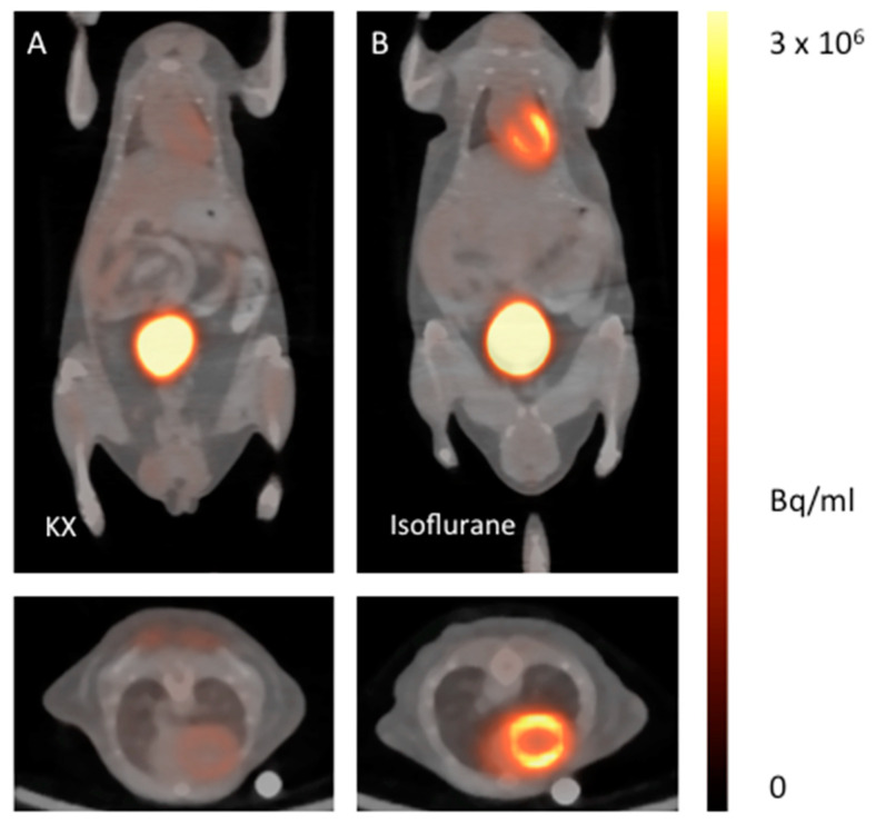
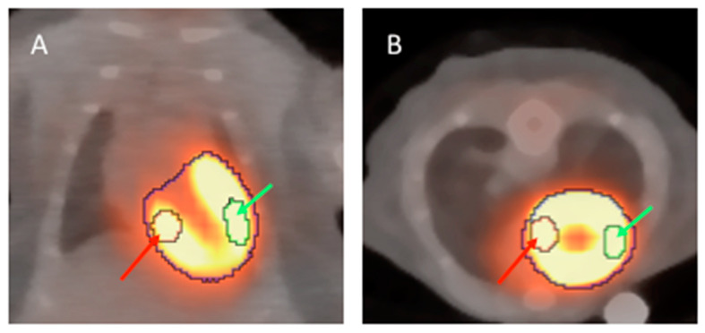
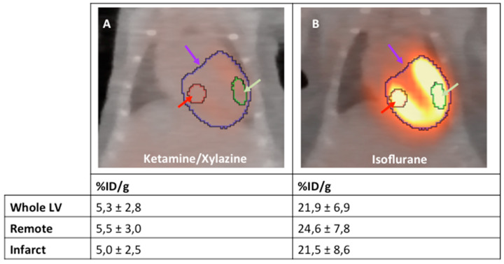
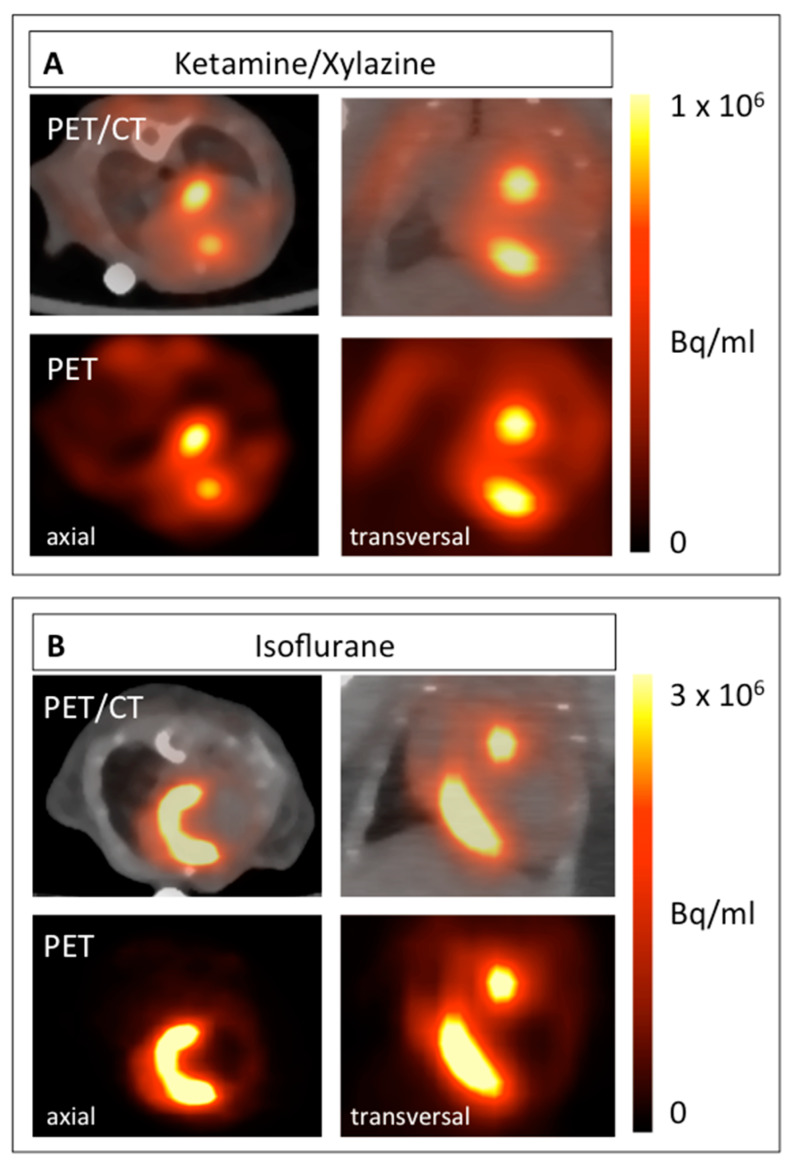
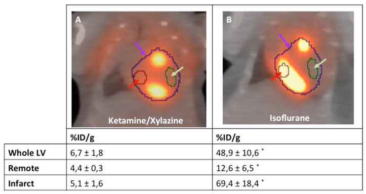
Cellular inflammation is an integral part of the healing process following acute myocardial infarction and has been under intense investigation for both therapeutic and prognostic approaches. Monocytes and macrophages are metabolically highly active and show increased uptake rates of glucose and its analog, F-FDG. Yet, the specific allocation of the radioactivity to the inflammatory cells via positron emission tomography (PET) imaging requires the suppression of glucose metabolism in viable myocardium. In mice, the most important model organism in basic research, this can be achieved by the application of ketamine/xylazine (KX) for anesthesia instead of isoflurane. Yet, while the consensus exists that glucose metabolism is effectively suppressed, a strategy for reproducible image analysis is grossly lacking and causes uncertainty concerning data interpretation. We introduce a simple strategy for systematic image analysis, which is a prerequisite to evaluate therapies targeting myocardial inflammation. Mice underwent permanent occlusion of the left anterior descending artery (LAD), inducing an acute myocardial infarction (MI). Five days after MI induction, 10MBq F-FDG was injected intravenously and a static PET/CT scan under ketamine/xylazine anesthesia was performed. For image reconstruction, we used an algorithm based on three-dimensional ordered subsets expectation maximization (3D-OSEM) followed by three-dimensional ordinary Poisson maximum a priori (MAP) reconstruction. Using this approach, high focal tracer uptake was typically located in the border zone of the infarct by visual inspection. To precisely demarcate the border zone for reproducible volume of interest (VOI) positioning, our protocol relies on positioning VOIs around the whole left ventricle, the inferobasal wall and the anterolateral wall guided by anatomical landmarks. This strategy enables comparable data in mouse studies, which is an important prerequisite for using a PET-based assessment of myocardial inflammation as a prognostic tool in therapeutic applications.
细胞炎症是急性心肌梗死后愈合过程的一个组成部分,一直是治疗和预后方法的研究热点。单核细胞和巨噬细胞代谢非常活跃,显示出葡萄糖及其类似物 F-FDG 的摄取率增加。然而,通过正电子发射断层扫描 (PET) 成像将放射性物质特异性分配给炎症细胞需要抑制存活心肌中的葡萄糖代谢。在基础研究中最重要的模式生物小鼠中,可以通过应用氯胺酮/二甲噻嗪 (KX) 麻醉代替异氟烷来实现。然而,尽管人们一致认为葡萄糖代谢得到了有效抑制,但缺乏可重复的图像分析策略,这导致对数据解释存在不确定性。我们介绍了一种简单的系统图像分析策略,这是评估针对心肌炎症的治疗方法的前提。小鼠接受左前降支 (LAD) 的永久性闭塞,诱导急性心肌梗死 (MI)。在 MI 诱导后 5 天,静脉注射 10MBq F-FDG,并在氯胺酮/二甲噻嗪麻醉下进行静态 PET/CT 扫描。对于图像重建,我们使用基于三维有序子集期望最大化 (3D-OSEM) 的算法,然后进行三维普通泊松最大后验 (MAP) 重建。使用这种方法,通过视觉检查,通常在梗塞的边界区域发现高焦点示踪剂摄取。为了精确地标定边界区域以进行可重复的感兴趣区 (VOI) 定位,我们的方案依赖于在解剖学标志物的引导下,围绕整个左心室、下基壁和前外侧壁定位 VOI,以重现 VOI 定位。这种策略能够在小鼠研究中获得可比的数据,这是将基于 PET 的心肌炎症评估作为治疗应用中的预后工具的重要前提。