Luo Yuxi, Qiao Mengyuan, Liang Yuqing, Chen Chongli, Zeng Lichuan, Wang Lin, Wu Wenbin
Department of Geriatrics, Hospital of Chengdu University of Traditional Chinese Medicine, Chengdu, China.
Department of Radiology, Hospital of Chengdu University of Traditional Chinese Medicine, Chengdu, China.
Front Aging Neurosci. 2022 Mar 10;14:812664. doi: 10.3389/fnagi.2022.812664. eCollection 2022.
To investigate the effect of sleep disorder (SD) on the changes of brain network dysfunction in mild cognitive impairment (MCI), we compared network connectivity patterns among MCI, SD, and comorbid MCI and sleep disorders (MCI-SD) patients using resting state functional magnetic resonance imaging (RS-fMRI).
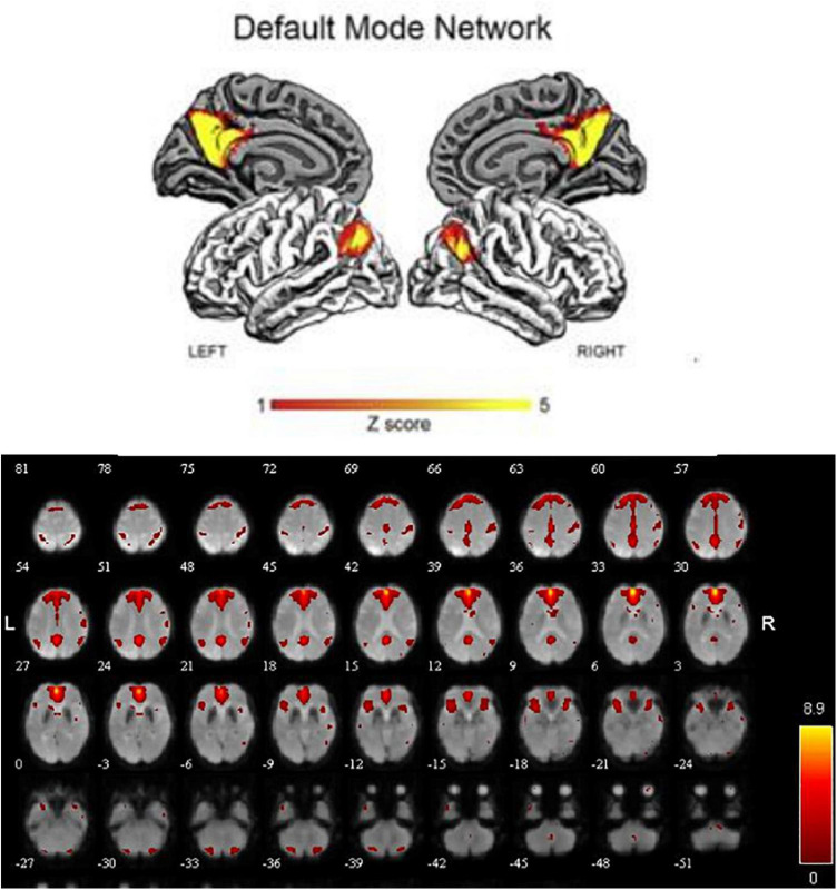
A total of 60 participants were included in this study, 20 each with MCI, SD, or MCI-SD. And all participants underwent structural and functional MRI scanning. The default-mode network (DMN) was extracted by independent component analysis (ICA), and regional functional connectivity strengths were calculated and compared among groups.
Compared to MCI patients, The DMN of MCI-SD patients demonstrated weaker functional connectivity with left middle frontal gyrus, right superior marginal gyrus, but stronger connectivity with the left parahippocampus, left precuneus and left middle temporal gyrus. Compared to the SD group, MCI-SD patients demonstrated weaker functional connectivity with right transverse temporal gyrus (Heschl's gyrus), right precentral gyrus, and left insula, but stronger connectivity with posterior cerebellum, right middle occipital gyrus, and left precuneus.
Patients with MCI-SD show unique changes in brain network connectivity patterns compared to MCI or SD alone, likely reflecting a broader functional disconnection and the need to recruit more brain regions for functional compensation.
为研究睡眠障碍(SD)对轻度认知障碍(MCI)患者脑网络功能障碍变化的影响,我们使用静息态功能磁共振成像(RS-fMRI)比较了MCI、SD以及合并MCI和睡眠障碍(MCI-SD)患者的网络连接模式。
本研究共纳入60名参与者,其中MCI、SD或MCI-SD患者各20名。所有参与者均接受了结构和功能MRI扫描。通过独立成分分析(ICA)提取默认模式网络(DMN),并计算组间区域功能连接强度并进行比较。
与MCI患者相比,MCI-SD患者的DMN与左侧额中回、右侧顶上小叶的功能连接较弱,但与左侧海马旁回、左侧楔前叶和左侧颞中回的连接较强。与SD组相比,MCI-SD患者与右侧颞横回(颞横回)、右侧中央前回和左侧岛叶的功能连接较弱,但与小脑后部、右侧枕中回和左侧楔前叶的连接较强。
与单独的MCI或SD相比,MCI-SD患者的脑网络连接模式有独特变化,可能反映了更广泛的功能断开以及需要调动更多脑区进行功能补偿。