Department of Biological Medicines & Shanghai Engineering Research Center of Immunotherapeutics, Fudan University School of Pharmacy, Shanghai, China.
Department of Chemistry, Fudan University, Shanghai, China.
Front Immunol. 2022 Mar 17;13:722053. doi: 10.3389/fimmu.2022.722053. eCollection 2022.
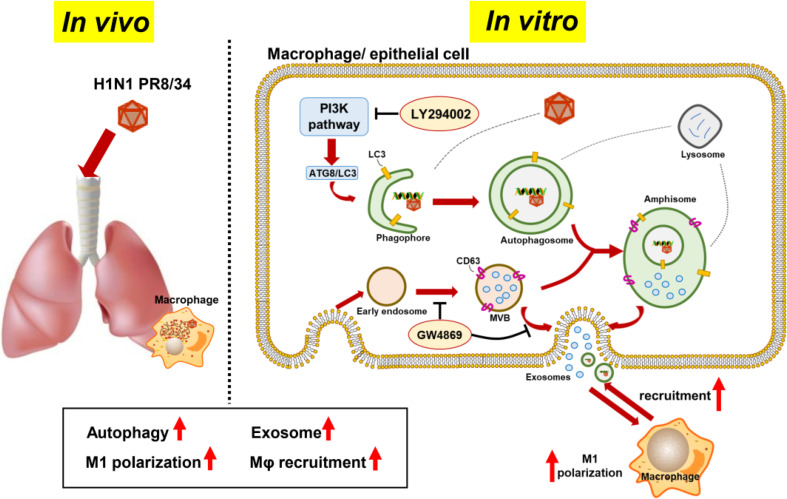
Influenza A virus infection results in viral pneumonia, which is often accompanied by the infiltration and recruitment of macrophages, overactivation of inflammatory responses, and obvious cell autophagy and exosome production. However, little is known about the roles of autophagy and exosome production in these inflammatory responses.
In this study, multiple methods, such as flow cytometry, real-time quantitative reverse transcription-polymerase chain reaction, immune-fluorescence technology, and western blot, were applied to explore the possible effects of autophagy and exosome production by H1N1-infected host cells.
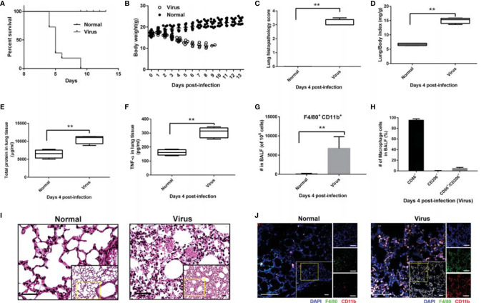
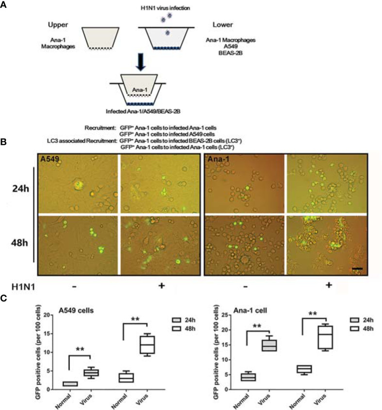
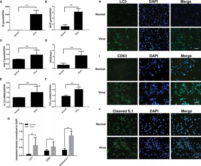
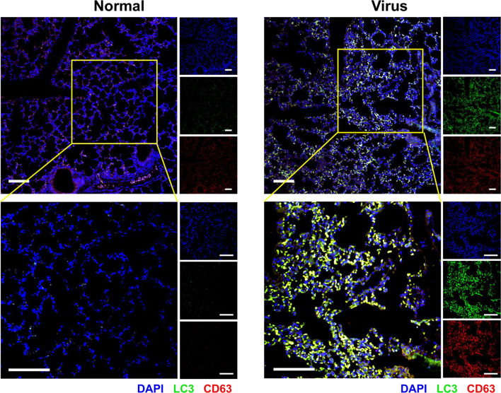
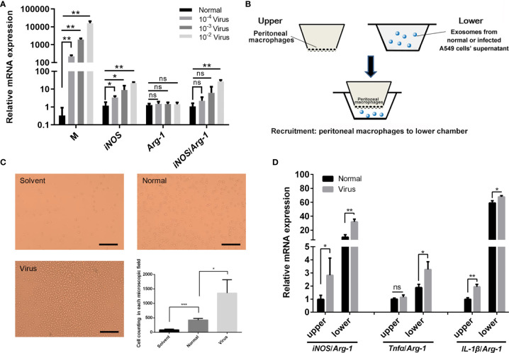
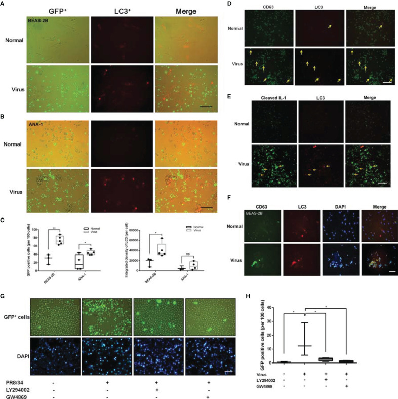
It was observed that a high number of polarized macrophages (CD11b/F4/80/CD86) were recruited to the lung tissues of infected mice, which could be mimicked by tracking the movement of macrophages to H1N1-infected cells (transwell assays). Furthermore, there was some coordinated upregulation of M1 polarization signs (iNOS/Arg-1 bias) as well as autophagy (LC3) and exosome (CD63) biomarkers in the infected macrophages and epithelial cells. Moreover, exosomes extracted from the supernatant of virus-infected cells were shown to promote the recruitment and polarization of more peritoneal macrophages than the normal group. The fluorescence colocalization of LC3-CD63 and the inhibition of autophagy and exosome signaling pathway further revealed that H1N1 infection seemed to sequentially activate the M1 polarization and recruitment of macrophages autophagy-exosome dependent pathway.
Autophagy and exosome production coordinately enhance the M1 polarization and recruitment of macrophages in influenza virus infection, which also provides potential therapeutic targets.
甲型流感病毒感染可导致病毒性肺炎,常伴有巨噬细胞浸润和募集、炎症反应过度激活以及明显的细胞自噬和外泌体产生。然而,对于自噬和外泌体产生在这些炎症反应中的作用知之甚少。
在这项研究中,应用流式细胞术、实时定量逆转录聚合酶链反应、免疫荧光技术和 Western blot 等多种方法,探讨了 H1N1 感染宿主细胞的自噬和外泌体产生的可能作用。
观察到大量极化的巨噬细胞(CD11b/F4/80/CD86)被招募到感染小鼠的肺组织中,通过追踪巨噬细胞向 H1N1 感染细胞(transwell 测定)的运动可以模拟这种现象。此外,在感染的巨噬细胞和上皮细胞中观察到 M1 极化标志(iNOS/Arg-1 偏向)以及自噬(LC3)和外泌体(CD63)生物标志物的协调上调。此外,从病毒感染细胞上清液中提取的外泌体被证明可促进更多腹腔巨噬细胞的募集和极化,而正常组则没有。LC3-CD63 的荧光共定位以及自噬和外泌体信号通路的抑制进一步表明,H1N1 感染似乎通过 M1 极化和依赖自噬-外泌体的巨噬细胞募集途径依次激活。
自噬和外泌体产生协同增强流感病毒感染中巨噬细胞的 M1 极化和募集,这也为潜在的治疗靶点提供了依据。