• 文献检索
  • 文档翻译
  • 深度研究
  • 学术资讯
  • Suppr Zotero 插件Zotero 插件
  • 邀请有礼
  • 套餐&价格
  • 历史记录
应用&插件
Suppr Zotero 插件Zotero 插件浏览器插件Mac 客户端Windows 客户端微信小程序
定价
高级版会员购买积分包购买API积分包
服务
文献检索文档翻译深度研究API 文档MCP 服务
关于我们
关于 Suppr公司介绍联系我们用户协议隐私条款
关注我们

Suppr 超能文献

核心技术专利:CN118964589B侵权必究
粤ICP备2023148730 号-1Suppr @ 2026

文献检索

告别复杂PubMed语法,用中文像聊天一样搜索,搜遍4000万医学文献。AI智能推荐,让科研检索更轻松。

立即免费搜索

文件翻译

保留排版,准确专业,支持PDF/Word/PPT等文件格式,支持 12+语言互译。

免费翻译文档

深度研究

AI帮你快速写综述,25分钟生成高质量综述,智能提取关键信息,辅助科研写作。

立即免费体验

应用超微血管成像测量的血管指数与乳腺癌分子亚型之间的关联

Association Between Vascular Index Measured Superb Microvascular Imaging and Molecular Subtype of Breast Cancer.

作者信息

Zhang Xiao-Yan, Cai Si-Man, Zhang Li, Zhu Qing-Li, Sun Qiang, Jiang Yu-Xin, Wang Hong-Yan, Li Jian-Chu

机构信息

Department of Diagnostic Ultrasound, Peking Union Medical College Hospital, Chinese Academy of Medical Sciences, Beijing, China.

Department of Breast Surgery, Peking Union Medical College Hospital, Chinese Academy of Medical Sciences, Beijing, China.

出版信息

Front Oncol. 2022 Mar 21;12:861151. doi: 10.3389/fonc.2022.861151. eCollection 2022.

DOI:10.3389/fonc.2022.861151
PMID:35387128
原文链接:https://pmc.ncbi.nlm.nih.gov/articles/PMC8979674/
Abstract

BACKGROUND

To determine whether vascular index (VI; defined as the ratio of Doppler signal pixels to pixels in the total lesion) measured superb microvascular imaging in breast cancer correlates with immunohistochemically defined subtype and is able to predict molecular subtypes.

METHODS

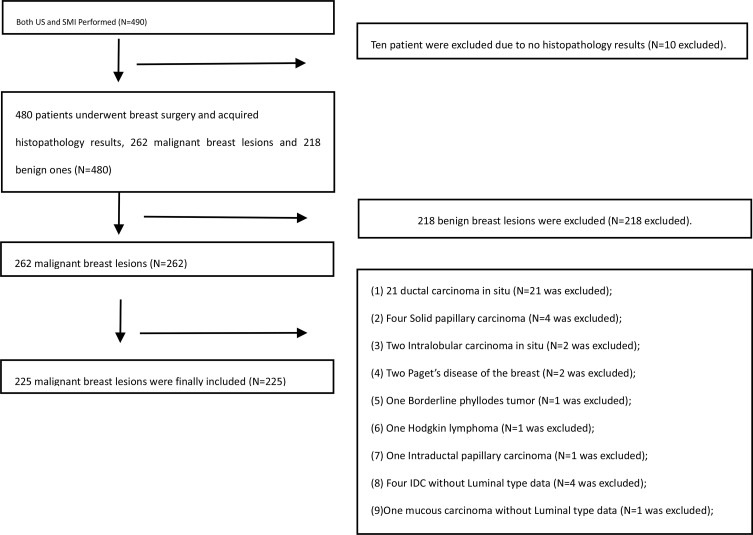
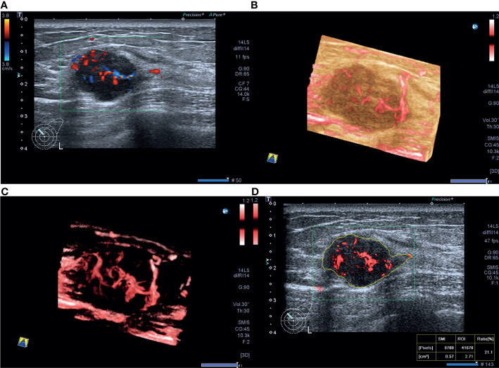
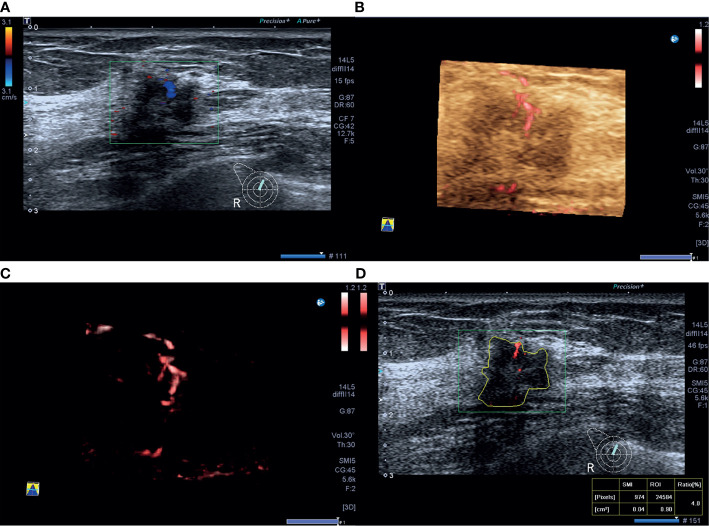
This prospective study involved 225 patients with 225 mass-type invasive breast cancers (mean size 2.6 ± 1.4 cm, range 0.4~5.9 cm) who underwent ultrasound and superb microvascular imaging (SMI) at Peking Union Medical College Hospital before breast surgery from December 2016 to June 2018. The correlations between primary tumor VI measured SMI, clinicopathological findings, and molecular subtype were analyzed. The performance of VI for prediction of molecular subtypes in invasive breast cancer was investigated.

RESULTS

The median VI of the 225 tumors was 7.3% (4.2%11.8%) (range 0%54.4%). Among the subtypes of the 225 tumors, 41 (18.2%) were luminal A, 91 (40.4%) were luminal B human epidermal growth factor receptor-2 (HER-2)-negative, 26 (11.6%) were luminal B HER-2-positive, 17 (7.6%) were HER-2-positive, and 50 (22.2%) were triple-negative, and the corresponding median VI values were 5.9% (2.6%11.6%) (range 0%47.1%), 7.3 (4.4%10.5%) (range 0%29.5%), 6.3% (3.9%11.3%) (range 0.6%22.2%), 8.2% (4.9%15.6%) (range 0.9%54.4%), and 9.2% (5.1%15.3%) (range 0.7%32.9%), respectively. Estrogen receptor (ER) negativity, higher tumor grade, and higher Ki-67 index (≥20%) were significantly associated with a higher VI value. Tumor size, ER status, and Ki-67 index were shown to independently influence VI. A cutoff value of 4.1% yielded 79.9% sensitivity and 41.5% specificity with an area under the receiver operating characteristic curve (AUC) of 0.58 for predicting that a tumor was of the luminal A subtype. A cutoff value of 16.4% yielded 30.0% sensitivity and 90.3% specificity with an AUC of 0.60 for predicting a triple-negative subtype.

CONCLUSIONS

VI, as a quantitative index obtained by SMI examination, could reflect histologic vascular changes in invasive breast cancer and was found to be higher in more biologically aggressive breast tumors. VI shows a certain degree of correlation with the molecular subtype of invasive breast cancer and plays a limited role in predicting the luminal A with high sensitivity and triple-negative subtype with high specificity.

摘要

背景

确定在乳腺癌中通过超声造影微血管成像测量的血管指数(VI,定义为多普勒信号像素与整个病变像素之比)是否与免疫组化定义的亚型相关,并能够预测分子亚型。

方法

这项前瞻性研究纳入了225例患有225个肿块型浸润性乳腺癌的患者(平均大小2.6±1.4 cm,范围0.4~5.9 cm),这些患者于2016年12月至2018年6月在北京协和医院接受乳房手术前接受了超声和超声造影微血管成像(SMI)检查。分析了通过SMI测量的原发性肿瘤VI、临床病理特征和分子亚型之间的相关性。研究了VI在预测浸润性乳腺癌分子亚型方面的性能。

结果

225个肿瘤的VI中位数为7.3%(4.2%11.8%)(范围0%54.4%)。在225个肿瘤的亚型中,41个(18.2%)为腔面A型,91个(40.4%)为腔面B型人表皮生长因子受体2(HER-2)阴性,26个(11.6%)为腔面B型HER-2阳性,17个(7.6%)为HER-2阳性,50个(22.2%)为三阴性,相应的VI中位数分别为5.9%(2.6%11.6%)(范围0%47.1%)、7.3(4.4%10.5%)(范围0%29.5%)、6.3%(3.9%11.3%)(范围 .6%22.2%)、8.2%(4.9%15.6%)(范围0.9%54.4%)和9.2%(5.1%15.3%)(范围0.7%32.9%)。雌激素受体(ER)阴性、更高的肿瘤分级和更高的Ki-67指数(≥20%)与更高的VI值显著相关。肿瘤大小、ER状态和Ki-67指数被证明独立影响VI。预测肿瘤为腔面A型时,截断值为4.1%时敏感性为79.9%,特异性为41.5%,受试者工作特征曲线(AUC)下面积为0.58。预测三阴性亚型时,截断值为16.4%时敏感性为30.0%,特异性为90.3%,AUC为0.60。

结论

VI作为通过SMI检查获得的定量指标,可以反映浸润性乳腺癌的组织学血管变化,并且在生物学行为更具侵袭性的乳腺肿瘤中更高。VI与浸润性乳腺癌的分子亚型显示出一定程度的相关性,在高敏感性预测腔面A型和高特异性预测三阴性亚型方面发挥有限作用。

https://cdn.ncbi.nlm.nih.gov/pmc/blobs/430f/8979674/42989a2d7943/fonc-12-861151-g003.jpg
https://cdn.ncbi.nlm.nih.gov/pmc/blobs/430f/8979674/39509aa91905/fonc-12-861151-g001.jpg
https://cdn.ncbi.nlm.nih.gov/pmc/blobs/430f/8979674/96b0a8167f81/fonc-12-861151-g002.jpg
https://cdn.ncbi.nlm.nih.gov/pmc/blobs/430f/8979674/42989a2d7943/fonc-12-861151-g003.jpg
https://cdn.ncbi.nlm.nih.gov/pmc/blobs/430f/8979674/39509aa91905/fonc-12-861151-g001.jpg
https://cdn.ncbi.nlm.nih.gov/pmc/blobs/430f/8979674/96b0a8167f81/fonc-12-861151-g002.jpg
https://cdn.ncbi.nlm.nih.gov/pmc/blobs/430f/8979674/42989a2d7943/fonc-12-861151-g003.jpg

相似文献

1
Association Between Vascular Index Measured Superb Microvascular Imaging and Molecular Subtype of Breast Cancer.应用超微血管成像测量的血管指数与乳腺癌分子亚型之间的关联
Front Oncol. 2022 Mar 21;12:861151. doi: 10.3389/fonc.2022.861151. eCollection 2022.
2
Prediction of Molecular Subtypes Using Superb Microvascular Imaging and Shear Wave Elastography in Invasive Breast Carcinomas.利用 Superb 微血管成像和剪切波弹性成像预测浸润性乳腺癌的分子亚型。
Acad Radiol. 2023 Jan;30(1):14-21. doi: 10.1016/j.acra.2022.04.017. Epub 2022 Jun 3.
3
Vascular index measured by smart 3-D superb microvascular imaging can help to differentiate malignant and benign breast lesion.通过智能三维超微血管成像测量的血管指数有助于鉴别乳腺良恶性病变。
Cancer Manag Res. 2019 Jun 13;11:5481-5487. doi: 10.2147/CMAR.S203376. eCollection 2019.
4
Association between ¹⁸F-FDG uptake and molecular subtype of breast cancer.¹⁸F-FDG摄取与乳腺癌分子亚型之间的关联。
Eur J Nucl Med Mol Imaging. 2015 Aug;42(9):1371-7. doi: 10.1007/s00259-015-3070-1. Epub 2015 May 7.
5
Added Value of the Vascular Index on Superb Microvascular Imaging for the Evaluation of Breast Masses: Comparison With Grayscale Ultrasound.超微血管成像的血管指数在乳腺肿块评估中的附加价值:与灰阶超声的比较
J Ultrasound Med. 2021 Apr;40(4):715-723. doi: 10.1002/jum.15441. Epub 2020 Aug 20.
6
[Application of superb microvascular imaging in focal liver lesions].[超微血管成像在肝脏局灶性病变中的应用]
Zhonghua Zhong Liu Za Zhi. 2021 Sep 23;43(9):959-967. doi: 10.3760/cma.j.cn112152-20191008-00649.
7
Reproducibility and diagnostic performance of the vascular index of superb microvascular imaging in real-time breast ultrasonography for evaluating breast masses.实时乳腺超声检查中用于评估乳腺肿块的超微血管成像血管指数的可重复性及诊断性能
Ultrasonography. 2021 Jul;40(3):398-406. doi: 10.14366/usg.20153. Epub 2020 Nov 27.
8
Long-Term Outcomes of Immunohistochemically Defined Subtypes of Breast Cancer Less Than or Equal to 2 cm After Breast-Conserving Surgery.保乳手术后最大直径小于或等于 2cm 的乳腺癌免疫组化定义亚型的长期预后
J Surg Res. 2019 Apr;236:288-299. doi: 10.1016/j.jss.2018.11.028. Epub 2018 Dec 27.
9
Journal Club: Diagnostic value of (18)F-FDG PET/CT and MRI in predicting the clinicopathologic subtypes of invasive breast cancer.期刊俱乐部:(18)F-FDG PET/CT 和 MRI 在预测浸润性乳腺癌临床病理亚型中的诊断价值。
AJR Am J Roentgenol. 2014 Aug;203(2):272-9. doi: 10.2214/AJR.13.11971.
10
Difference between Luminal A and Luminal B Subtypes According to Ki-67, Tumor Size, and Progesterone Receptor Negativity Providing Prognostic Information.根据 Ki-67、肿瘤大小和孕激素受体阴性对 Luminal A 和 Luminal B 亚型进行区分,提供预后信息。
Clin Med Insights Oncol. 2014 Sep 11;8:107-11. doi: 10.4137/CMO.S18006. eCollection 2014.

引用本文的文献

1
AI-assisted diffuse correlation tomography for identifying breast cancer.用于识别乳腺癌的人工智能辅助扩散相关断层扫描技术
J Biomed Opt. 2025 May;30(5):055001. doi: 10.1117/1.JBO.30.5.055001. Epub 2025 May 16.
2
Pilot Study to Evaluate the Association Between Superb Microvascular Imaging (SMI) and Histologic Markers of Angiogenesis in Patients With Invasive Ductal Carcinoma.评估超声微血管成像(SMI)与浸润性导管癌患者血管生成组织学标志物之间关联的初步研究。
J Ultrasound Med. 2025 Jul;44(7):1201-1211. doi: 10.1002/jum.16674. Epub 2025 Mar 5.
3
Role of Superb Microvascular Imaging (SMI) vascularity index values and vascularity patterns in the differential diagnosis of malignant liver lesions.

本文引用的文献

1
Comparison of the Effectiveness of Shear Wave Elastography and Superb Microvascular Imaging in the Evaluation of Breast Masses.剪切波弹性成像与超微血管成像在乳腺肿块评估中的效果比较。
Ultrasound Q. 2021 Jun 1;37(2):191-197. doi: 10.1097/RUQ.0000000000000562.
2
Reproducibility and diagnostic performance of the vascular index of superb microvascular imaging in real-time breast ultrasonography for evaluating breast masses.实时乳腺超声检查中用于评估乳腺肿块的超微血管成像血管指数的可重复性及诊断性能
Ultrasonography. 2021 Jul;40(3):398-406. doi: 10.14366/usg.20153. Epub 2020 Nov 27.
3
Added Value of the Vascular Index on Superb Microvascular Imaging for the Evaluation of Breast Masses: Comparison With Grayscale Ultrasound.
超微血管成像(SMI)血管指数值和血管模式在肝脏恶性病变鉴别诊断中的作用
Abdom Radiol (NY). 2025 Jun;50(6):2426-2439. doi: 10.1007/s00261-024-04711-z. Epub 2024 Nov 22.
4
From Chaos to Opportunity: Decoding Cancer Heterogeneity for Enhanced Treatment Strategies.从混乱到机遇:解读癌症异质性以优化治疗策略
Biology (Basel). 2023 Aug 29;12(9):1183. doi: 10.3390/biology12091183.
5
Combining conventional ultrasound and ultrasound elastography to predict HER2 status in patients with breast cancer.联合传统超声与超声弹性成像预测乳腺癌患者的人表皮生长因子受体2(HER2)状态
Front Physiol. 2023 Jul 12;14:1188502. doi: 10.3389/fphys.2023.1188502. eCollection 2023.
6
Conventional ultrasound and contrast-enhanced ultrasound radiomics in breast cancer and molecular subtype diagnosis.传统超声和超声造影在乳腺癌及分子亚型诊断中的影像组学研究
Front Oncol. 2023 May 23;13:1158736. doi: 10.3389/fonc.2023.1158736. eCollection 2023.
7
Volumetric imaging and morphometric analysis of breast tumor angiogenesis using a new contrast-free ultrasound technique: a feasibility study.利用一种新的无对比超声技术对乳腺肿瘤血管生成进行容积成像和形态计量分析:一项可行性研究。
Breast Cancer Res. 2022 Nov 30;24(1):85. doi: 10.1186/s13058-022-01583-3.
超微血管成像的血管指数在乳腺肿块评估中的附加价值:与灰阶超声的比较
J Ultrasound Med. 2021 Apr;40(4):715-723. doi: 10.1002/jum.15441. Epub 2020 Aug 20.
4
Combination of Quantitative Parameters of Shear Wave Elastography and Superb Microvascular Imaging to Evaluate Breast Masses.剪切波弹性成像定量参数与超微血管成像联合评估乳腺肿块。
Korean J Radiol. 2020 Sep;21(9):1045-1054. doi: 10.3348/kjr.2019.0765.
5
Correlation between imaging features and molecular subtypes of breast cancer in young women (≤30 years old).年轻女性(≤30岁)乳腺癌的影像学特征与分子亚型之间的相关性
Jpn J Radiol. 2020 Nov;38(11):1062-1074. doi: 10.1007/s11604-020-01001-8. Epub 2020 Jun 19.
6
FOXA1 upregulation promotes enhancer and transcriptional reprogramming in endocrine-resistant breast cancer.FOXA1上调促进内分泌抵抗性乳腺癌中的增强子和转录重编程。
Proc Natl Acad Sci U S A. 2019 Dec 26;116(52):26823-26834. doi: 10.1073/pnas.1911584116. Epub 2019 Dec 11.
7
Breast cancer statistics, 2019.乳腺癌统计数据,2019 年。
CA Cancer J Clin. 2019 Nov;69(6):438-451. doi: 10.3322/caac.21583. Epub 2019 Oct 2.
8
Vascular index measured by smart 3-D superb microvascular imaging can help to differentiate malignant and benign breast lesion.通过智能三维超微血管成像测量的血管指数有助于鉴别乳腺良恶性病变。
Cancer Manag Res. 2019 Jun 13;11:5481-5487. doi: 10.2147/CMAR.S203376. eCollection 2019.
9
A Prospective Study on the Value of Ultrasound Microflow Assessment to Distinguish Malignant from Benign Solid Breast Masses: Association between Ultrasound Parameters and Histologic Microvessel Densities.超声微血管流评估对鉴别良恶性实性乳腺肿块的价值的前瞻性研究:超声参数与组织微血管密度的相关性。
Korean J Radiol. 2019 May;20(5):759-772. doi: 10.3348/kjr.2018.0515.
10
Ki-67 labeling index as a prognostic marker in advanced stomach cancer.Ki-67标记指数作为晚期胃癌的预后标志物
Ann Surg Treat Res. 2019 Jan;96(1):27-33. doi: 10.4174/astr.2019.96.1.27. Epub 2018 Dec 26.