Lee Eun Ji, Chang Yun-Woo, Oh Eunsun, Hwang Jiyoung, Kim Hyun-Joo, Hong Seong Sook
Department of Radiology, Soonchunhyang University Seoul Hospital, Seoul, Korea.
Ultrasonography. 2021 Jul;40(3):398-406. doi: 10.14366/usg.20153. Epub 2020 Nov 27.
This study aimed to evaluate the reproducibility and diagnostic performance of a quantitative parameter of superb microvascular imaging (SMI) in real-time breast ultrasonography (US) for differentiating benign from malignant breast masses.
Eighty-seven breast masses in 75 patients who underwent both B-mode US and SMI before US-guided core needle biopsy were included in this study. Two radiologists performed B-mode US and measured the vascular index (VI) of SMI respectively for each lesion in real time. Intraobserver and interobserver agreements were analyzed for the VI of SMI. The diagnostic performance of B-mode US using the Breast Imaging Reporting and Database System lexicon and combined use with the VI of SMI was evaluated compared to pathology.
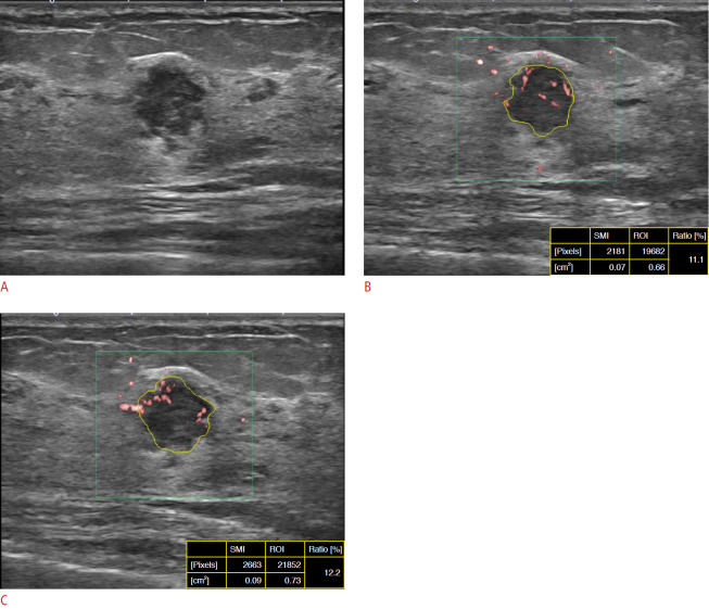
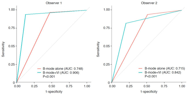
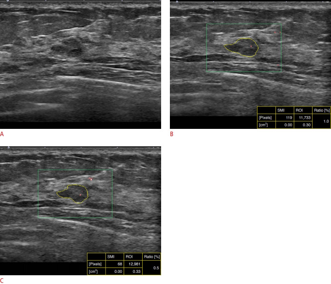
The median VI of malignant masses (n=32) was significantly higher than that of benign masses (n=55) (7.6% and 2.6%, respectively; P<0.001). The intraobserver agreement for VI was excellent regardless of the pathology, size, or depth of the lesion. The interobserver agreement for VI was excellent regardless of the presence of a measurement interval. The interobserver agreement for the final diagnostic decision was improved by combining B-mode US and VI (κ=0.883) in comparison with B-mode US only (κ=0.617). Adding VI led to significant improvements in the specificity (87.2% vs. 52.7%, 83.6% vs. 49.0%), accuracy (89.7% vs. 69.3%, 84.0% vs. 65.9%) and positive predictive value (81.5% vs. 55.1%, 75.6% vs. 52.6%) of B-mode US for both observers compared with B-mode US alone (all, P=0.001).
The VI of SMI for real-time breast US is highly reproducible and leads to improved diagnostic performance for differentiating between benign and malignant breast lesions in combination with B-mode US.
本研究旨在评估实时乳腺超声检查中,超微血管成像(SMI)定量参数在鉴别乳腺良恶性肿块方面的可重复性及诊断性能。
本研究纳入了75例患者的87个乳腺肿块,这些患者在超声引导下进行粗针穿刺活检前均接受了B超和SMI检查。两名放射科医生分别进行B超检查,并实时测量每个病灶的SMI血管指数(VI)。分析了SMI的VI在观察者内和观察者间的一致性。与病理结果相比,评估了使用乳腺影像报告和数据系统词典的B超以及结合SMI的VI的诊断性能。
恶性肿块(n = 32)的VI中位数显著高于良性肿块(n = 55)(分别为7.6%和2.6%;P<0.001)。无论病灶的病理类型、大小或深度如何,VI的观察者内一致性均极佳。无论是否存在测量间隔,VI的观察者间一致性均极佳。与仅使用B超相比,联合使用B超和VI可提高观察者间最终诊断决策的一致性(κ=0.883),而仅使用B超的一致性为(κ=0.617)。与单独使用B超相比,添加VI可显著提高两名观察者的B超特异性(87.2%对52.7%,83.6%对49.0%)、准确性(89.7%对69.3%,84.0%对65.9%)和阳性预测值(81.5%对55.1%,75.6%对52.6%)(所有P=0.001)。
实时乳腺超声检查中SMI的VI具有高度可重复性,与B超联合使用可提高鉴别乳腺良恶性病变的诊断性能。