Joint Inspection Center of Precision Medicine, The People's Hospital of Guangxi Zhuang Autonomous Region & Guangxi Academy of Medical Sciences, Nanning, Guangxi, People's Republic of China.
Department of Clinical Laboratory, the first affiliated hospital of Guangxi University of Chinese Medicine, Nanning, Guangxi, People's Republic of China.
BMC Cancer. 2022 Apr 7;22(1):370. doi: 10.1186/s12885-022-09386-7.
T-cell immunoglobulin mucin-1 (TIM-1) has been reported to be associated with the biological behavior of several malignant tumors; however, it is not clear whether it has a role in cervical cancer (CC).
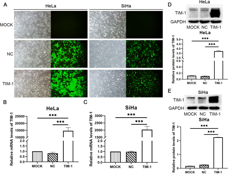
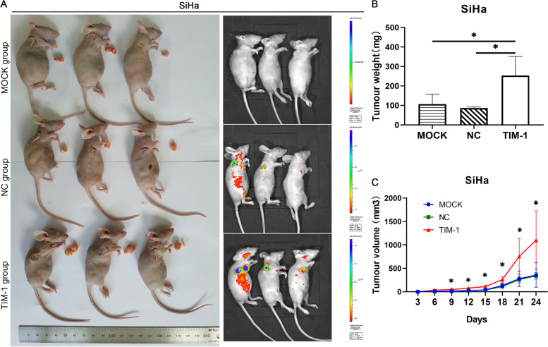
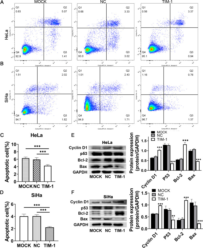
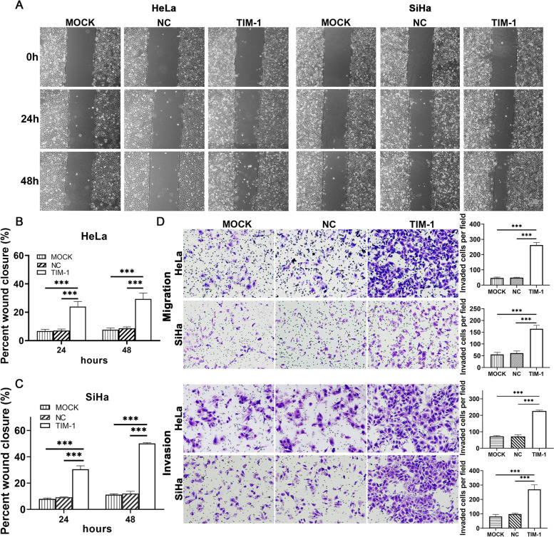
TIM-1 expression in cervical epithelial tumor tissues and cells was detected by immunohistochemistry or real-time quantitative-PCR and western blotting. CC cells from cell lines expressing low levels of TIM-1 were infected with lentiviral vectors encoding TIM-1. Changes in the malignant behavior of CC cells were assessed by CCK-8, wound healing, Transwell migration and invasion assays, and flow cytometry in vitro; while a xenograft tumor model was established to analyze the effects of TIM-1 on tumor growth in vivo. Changes in the levels of proteins related to the cell cycle, apoptosis, and Epithelial-mesenchymal transition (EMT) were determined by western blotting.
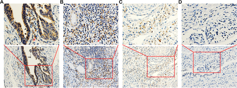
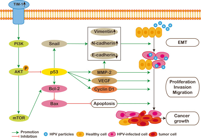
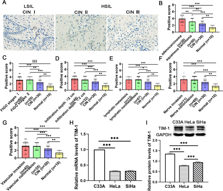
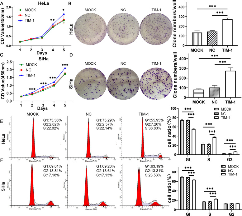
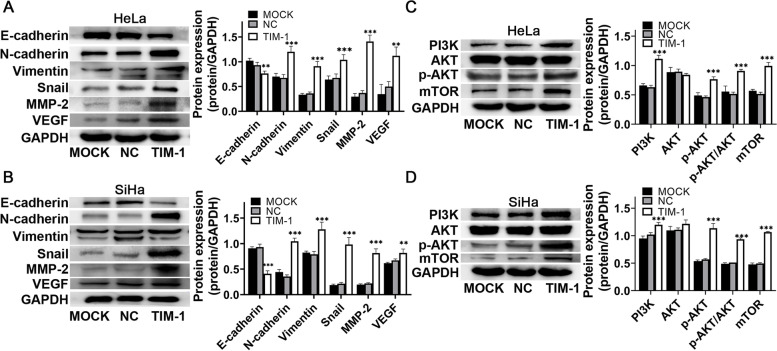
TIM-1 expression was higher in CC tissues, than in high grade squamous intraepithelial lesion, low grade squamous intraepithelial lesion, or normal cervical tissues, and was also expressed in three CC cell lines. In HeLa and SiHa cells overexpressing TIM-1, proliferation, invasion, and migration increased, while whereas apoptosis was inhibited. Furthermore, TIM-1 downregulated the expression of p53, BAX, and E-cadherin, and increased cyclin D1, Bcl-2, Snail1, N-cadherin, vimentin, MMP-2, and VEGF. PI3K, p-AKT, and mTOR protein levels also increased, while total AKT protein levels remained unchanged.
Our study indicated that TIM-1 overexpression promoted cell migration and invasion, and inhibited cell apoptosis in CC through modulation of the PI3K/AKT/p53 and PI3K/AKT/mTOR signaling pathways, and may be a candidate diagnostic biomarker of this disease.
T 细胞免疫球蛋白黏蛋白-1(TIM-1)已被报道与几种恶性肿瘤的生物学行为有关;然而,它在宫颈癌(CC)中的作用尚不清楚。
采用免疫组化或实时定量 PCR 和 Western blot 检测宫颈上皮肿瘤组织和细胞中 TIM-1 的表达。用慢病毒载体感染 TIM-1 低表达的 CC 细胞系,以沉默 TIM-1 的表达。体外通过 CCK-8、划痕愈合、Transwell 迁移和侵袭实验以及流式细胞术评估 CC 细胞恶性行为的变化;同时建立异种移植肿瘤模型,分析 TIM-1 对体内肿瘤生长的影响。通过 Western blot 测定与细胞周期、凋亡和上皮间质转化(EMT)相关的蛋白水平变化。
CC 组织中 TIM-1 的表达高于高级别鳞状上皮内病变、低级别鳞状上皮内病变或正常宫颈组织,且在三种 CC 细胞系中也有表达。在 TIM-1 过表达的 HeLa 和 SiHa 细胞中,增殖、侵袭和迁移增加,而凋亡受到抑制。此外,TIM-1 下调了 p53、BAX 和 E-cadherin 的表达,上调了 cyclin D1、Bcl-2、Snail1、N-cadherin、vimentin、MMP-2 和 VEGF 的表达。PI3K、p-AKT 和 mTOR 蛋白水平也升高,而总 AKT 蛋白水平保持不变。
我们的研究表明,TIM-1 过表达通过调节 PI3K/AKT/p53 和 PI3K/AKT/mTOR 信号通路促进 CC 细胞迁移和侵袭,并抑制细胞凋亡,可能是该疾病的候选诊断生物标志物。