• 文献检索
  • 文档翻译
  • 深度研究
  • 学术资讯
  • Suppr Zotero 插件Zotero 插件
  • 邀请有礼
  • 套餐&价格
  • 历史记录
应用&插件
Suppr Zotero 插件Zotero 插件浏览器插件Mac 客户端Windows 客户端微信小程序
定价
高级版会员购买积分包购买API积分包
服务
文献检索文档翻译深度研究API 文档MCP 服务
关于我们
关于 Suppr公司介绍联系我们用户协议隐私条款
关注我们

Suppr 超能文献

核心技术专利:CN118964589B侵权必究
粤ICP备2023148730 号-1Suppr @ 2026

文献检索

告别复杂PubMed语法,用中文像聊天一样搜索,搜遍4000万医学文献。AI智能推荐,让科研检索更轻松。

立即免费搜索

文件翻译

保留排版,准确专业,支持PDF/Word/PPT等文件格式,支持 12+语言互译。

免费翻译文档

深度研究

AI帮你快速写综述,25分钟生成高质量综述,智能提取关键信息,辅助科研写作。

立即免费体验

下颌头的骨合成,螺钉的放置方式重要吗?

The Osteosynthesis of the Mandibular Head, Does the Way the Screws Are Positioned Matter?

作者信息

Kozakiewicz Marcin, Gabryelczak Izabela

机构信息

Department of Maxillofacial Surgery, Medical University of Lodz, 113 Żeromskiego Str., 90-549 Lodz, Poland.

出版信息

J Clin Med. 2022 Apr 5;11(7):2031. doi: 10.3390/jcm11072031.

DOI:10.3390/jcm11072031
PMID:35407637
原文链接:https://pmc.ncbi.nlm.nih.gov/articles/PMC8999701/
Abstract

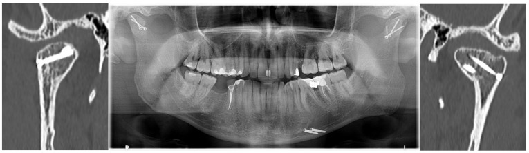
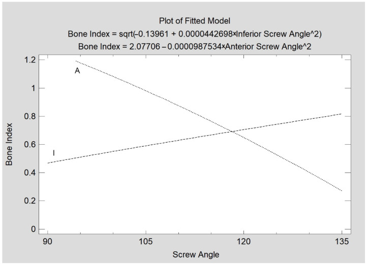
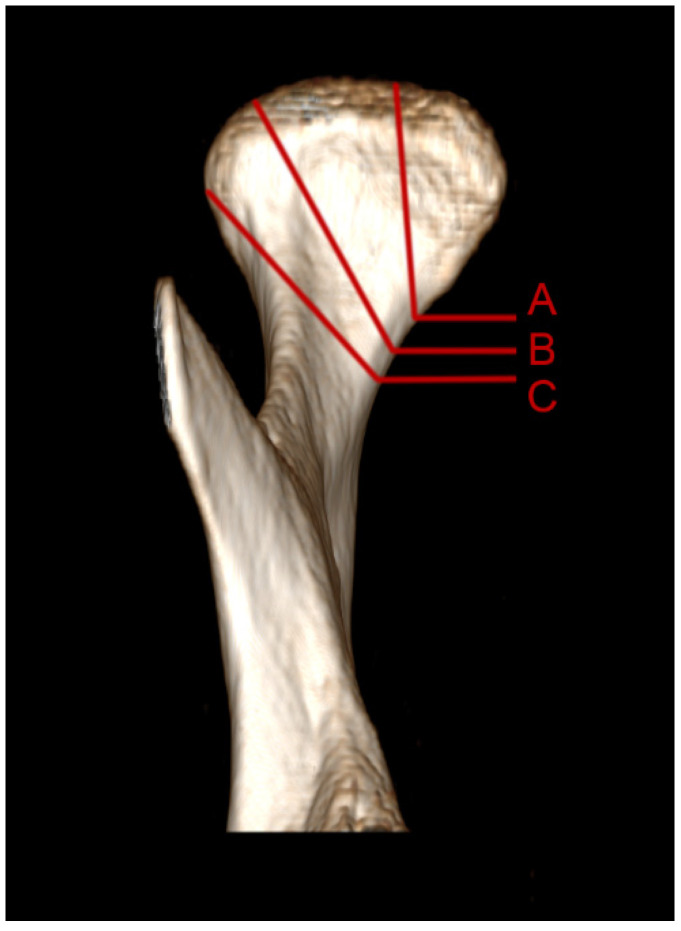
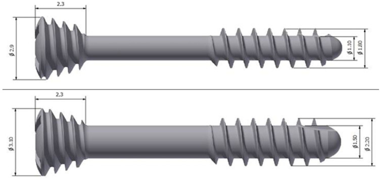
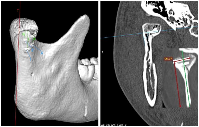
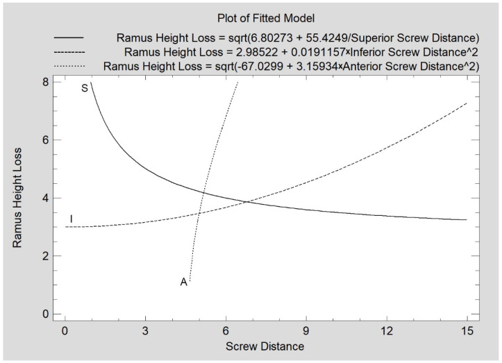
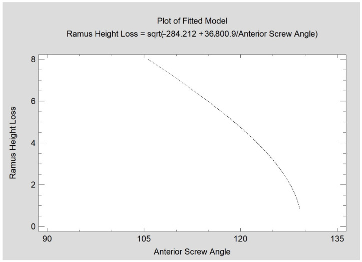
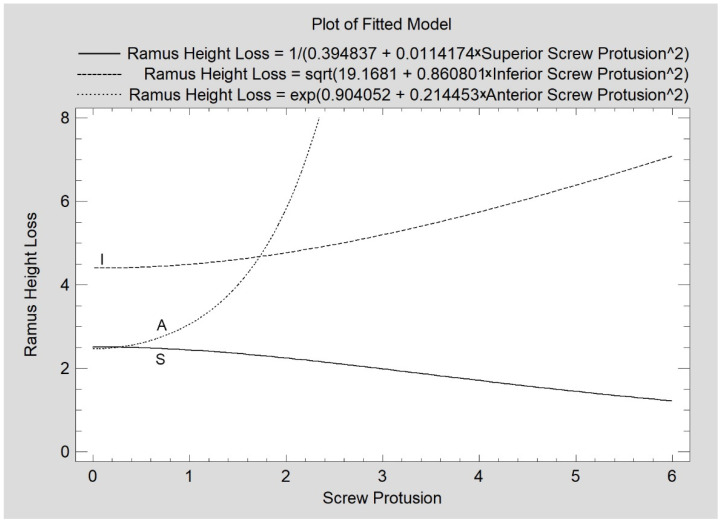
Currently, an increasing number of medical centers are treating mandibular head fractures surgically. Dedicated screws for compression osteosynthesis have been developed. However, due to the very limited size of the fractured bones and the considerable technical difficulties accompanying the execution of the fixation, there is little room for correction of the positioning and reinsertion of the screws. Therefore, knowing the optimal position of the fixation material is crucial for therapeutic success. The aim of this study is the evaluation of fixation screw position on the mandibular ramus height obtained in the treatment of the condylar head fracture. A total of 57 patients were included in this study. The loss of mandibular ramus height on computed tomography twelve months after mandibular head osteosynthesis was evaluated in relation to the initial distance of the screws from the fracture line, the angle of insertion of the screw into the bone, and the size of the protrusion to the inner side of the condyle. The relationship of the proximity of the screw to the fracture line, angulation, and the size of the protrusion with the loss of ramus height was confirmed. Conclusions: the optimal location for the superior screw is approx. 4 mm below the fracture line (with any angulation), inferior screw is approx. 8 mm (with any angulation), and anterior screw position is approx. 4-5 mm distant from fracture line with the best angulation of 130 degrees to the lateral mandible ramus surface in the coronal plane.

摘要

目前,越来越多的医疗中心采用手术治疗下颌骨髁突骨折。专门用于加压接骨术的螺钉已被研发出来。然而,由于骨折部位的骨头尺寸非常有限,且在进行固定操作时存在相当大的技术困难,螺钉定位调整和重新插入的空间很小。因此,了解固定材料的最佳位置对于治疗成功至关重要。本研究的目的是评估在髁突骨折治疗中获得的下颌支高度上固定螺钉的位置。本研究共纳入57例患者。根据螺钉与骨折线的初始距离、螺钉插入骨头的角度以及髁突内侧突出的大小,评估下颌骨髁突接骨术后十二个月计算机断层扫描中下颌支高度的丢失情况。证实了螺钉与骨折线的接近程度、角度以及突出大小与下颌支高度丢失之间的关系。结论:上方螺钉的最佳位置约在骨折线下方4毫米处(任意角度),下方螺钉约为8毫米(任意角度),前方螺钉位置距骨折线约4 - 5毫米,在冠状面与下颌支外侧表面的最佳角度为130度。

https://cdn.ncbi.nlm.nih.gov/pmc/blobs/a048/8999701/72bef8995615/jcm-11-02031-g009.jpg
https://cdn.ncbi.nlm.nih.gov/pmc/blobs/a048/8999701/42918aed91f4/jcm-11-02031-g001.jpg
https://cdn.ncbi.nlm.nih.gov/pmc/blobs/a048/8999701/a6e12c80b652/jcm-11-02031-g002.jpg
https://cdn.ncbi.nlm.nih.gov/pmc/blobs/a048/8999701/9eabfc66c74f/jcm-11-02031-g003.jpg
https://cdn.ncbi.nlm.nih.gov/pmc/blobs/a048/8999701/910d846ea270/jcm-11-02031-g004.jpg
https://cdn.ncbi.nlm.nih.gov/pmc/blobs/a048/8999701/78e41c1fda10/jcm-11-02031-g005.jpg
https://cdn.ncbi.nlm.nih.gov/pmc/blobs/a048/8999701/9139e81cb5fb/jcm-11-02031-g006.jpg
https://cdn.ncbi.nlm.nih.gov/pmc/blobs/a048/8999701/a7242d1fe3f7/jcm-11-02031-g007.jpg
https://cdn.ncbi.nlm.nih.gov/pmc/blobs/a048/8999701/6c31fbe8bf04/jcm-11-02031-g008.jpg
https://cdn.ncbi.nlm.nih.gov/pmc/blobs/a048/8999701/72bef8995615/jcm-11-02031-g009.jpg

相似文献

1
The Osteosynthesis of the Mandibular Head, Does the Way the Screws Are Positioned Matter?下颌头的骨合成,螺钉的放置方式重要吗?
J Clin Med. 2022 Apr 5;11(7):2031. doi: 10.3390/jcm11072031.
2
Clinical Evaluation of Magnesium Alloy Osteosynthesis in the Mandibular Head.镁合金在下颌骨髁突骨合成中的临床评估
Materials (Basel). 2022 Jan 18;15(3):711. doi: 10.3390/ma15030711.
3
Two versus three magnesium screws for osteosynthesis of mandibular condylar head fractures: a finite element analysis.两种与三种用于下颌骨髁突骨折内固定的镁螺钉:有限元分析。
Clin Oral Investig. 2024 Sep 27;28(10):553. doi: 10.1007/s00784-024-05927-5.
4
Bone Union Quality after Fracture Fixation of Mandibular Head with Compression Magnesium Screws.使用加压镁螺钉固定下颌头骨折后的骨愈合质量
Materials (Basel). 2022 Mar 17;15(6):2230. doi: 10.3390/ma15062230.
5
Finite element analysis of type B condylar head fractures and osteosynthesis using two positional screws.使用两枚定位螺钉对B型髁状头骨折及骨内固定术的有限元分析
J Craniomaxillofac Surg. 2014 Jul;42(5):482-8. doi: 10.1016/j.jcms.2013.06.006. Epub 2013 Jul 30.
6
Comparison of compression screws used for mandible head fracture treatment-experimental study.用于下颌骨骨折治疗的压缩螺钉的比较-实验研究。
Clin Oral Investig. 2019 Nov;23(11):4059-4066. doi: 10.1007/s00784-019-02842-y. Epub 2019 Mar 20.
7
Two-screw osteosynthesis of the mandibular condylar head with different screw materials: a finite element analysis.不同螺钉材料的下颌髁突双螺钉固定:有限元分析。
Comput Methods Biomech Biomed Engin. 2024 May;27(7):878-882. doi: 10.1080/10255842.2023.2209247. Epub 2023 May 8.
8
Treatment of mandibular-condylar fractures.下颌髁突骨折的治疗。
Atlas Oral Maxillofac Surg Clin North Am. 1997 Mar;5(1):127-55.
9
Long-term results of ORIF of condylar head fractures of the mandible: A prospective 5-year follow-up study of small-fragment positional-screw osteosynthesis (SFPSO).下颌骨髁突头部骨折切开复位内固定术的长期结果:小碎片定位螺钉骨合成术(SFPSO)的前瞻性5年随访研究
J Craniomaxillofac Surg. 2015 May;43(4):452-61. doi: 10.1016/j.jcms.2015.02.004. Epub 2015 Feb 13.
10
Two-versus three-screw osteosynthesis of the mandibular condylar head: A finite element analysis.双螺钉与三螺钉下颌骨髁突骨固定术的有限元分析。
J Mech Behav Biomed Mater. 2022 Mar;127:105077. doi: 10.1016/j.jmbbm.2022.105077. Epub 2022 Jan 8.

引用本文的文献

1
Risk of Permanent Dysfunction of Facial Nerves After Open Rigid Internal Fixation in the Treatment of Mandibular Condylar Process Fracture.下颌髁突骨折切开坚固内固定术后面神经永久性功能障碍的风险
Med Sci (Basel). 2025 Aug 9;13(3):121. doi: 10.3390/medsci13030121.
2
Fixation of Medially Displaced High Condylar Fracture of Mandible Using Lag Screw: A Twin Case Report.使用拉力螺钉固定下颌骨内侧移位髁突高位骨折:一例双病例报告
J Maxillofac Oral Surg. 2025 Apr;24(2):401-405. doi: 10.1007/s12663-024-02206-4. Epub 2024 May 30.
3
Measures of Corticalization.

本文引用的文献

1
Bone Union Quality after Fracture Fixation of Mandibular Head with Compression Magnesium Screws.使用加压镁螺钉固定下颌头骨折后的骨愈合质量
Materials (Basel). 2022 Mar 17;15(6):2230. doi: 10.3390/ma15062230.
2
Clinical Evaluation of Magnesium Alloy Osteosynthesis in the Mandibular Head.镁合金在下颌骨髁突骨合成中的临床评估
Materials (Basel). 2022 Jan 18;15(3):711. doi: 10.3390/ma15030711.
3
Are recent available blended collagen-calcium phosphate better than collagen alone or crystalline calcium phosphate? Radiotextural analysis of a 1-year clinical trial.
皮质化测量
J Clin Med. 2022 Sep 16;11(18):5463. doi: 10.3390/jcm11185463.
4
Repeated Intra-Articular Administration of Platelet-Rich Plasma (PRP) in Temporomandibular Disorders: A Clinical Case Series.富血小板血浆(PRP)在颞下颌关节紊乱病中的反复关节内注射:临床病例系列
J Clin Med. 2022 Jul 22;11(15):4281. doi: 10.3390/jcm11154281.
近期可用的混和胶原-磷酸钙是否优于单独胶原或结晶磷酸钙?为期 1 年的临床试验的放射学分析。
Clin Oral Investig. 2021 Jun;25(6):3711-3718. doi: 10.1007/s00784-020-03697-4. Epub 2020 Nov 23.
4
Biomechanical evaluation of type p condylar head osteosynthesis using conventional small-fragment screws reinforced by a patient specific two-component plate.采用患者特异性双组件接骨板增强的常规小片段螺钉对 p 型髁突头骨折进行生物力学评估。
Head Face Med. 2020 Oct 19;16(1):25. doi: 10.1186/s13005-020-00236-0.
5
Application of CAD/CAM technology for surgical treatment of condylar head fractures: A preliminary study.计算机辅助设计/计算机辅助制造(CAD/CAM)技术在髁突头部骨折手术治疗中的应用:一项初步研究。
J Oral Biol Craniofac Res. 2020 Oct-Dec;10(4):608-614. doi: 10.1016/j.jobcr.2020.08.018. Epub 2020 Aug 27.
6
Influence of General Mineral Condition on Collagen-Guided Alveolar Crest Augmentation.一般矿物质状况对胶原引导的牙槽嵴增高的影响。
Materials (Basel). 2020 Aug 18;13(16):3649. doi: 10.3390/ma13163649.
7
Preauricular Swelling Mimicking a Tumour: Dissolution of Mandibular Capitulum Following Trauma in a 15-Year Old Child.前耳肿胀酷似肿瘤:15 岁儿童外伤后下颌骨小头溶解。
In Vivo. 2020 May-Jun;34(3):1235-1245. doi: 10.21873/invivo.11897.
8
Nonunion - consensus from the 4th annual meeting of the Danish Orthopaedic Trauma Society.骨不连——丹麦骨创伤学会第四届年会共识
EFORT Open Rev. 2020 Jan 29;5(1):46-57. doi: 10.1302/2058-5241.5.190037. eCollection 2020 Jan.
9
Key points in surgical management of mandibular condylar fractures.下颌髁突骨折手术治疗的要点
Chin J Traumatol. 2020 Apr;23(2):63-70. doi: 10.1016/j.cjtee.2019.08.006. Epub 2019 Nov 1.
10
The application of virtual reality and augmented reality in Oral & Maxillofacial Surgery.虚拟现实和增强现实在口腔颌面外科学中的应用。
BMC Oral Health. 2019 Nov 8;19(1):238. doi: 10.1186/s12903-019-0937-8.